Andhra Pradesh BIEAP AP Inter 2nd Year Zoology Study Material Lesson 2(b) Excretory Products and their Elimination Textbook Questions and Answers.
AP Inter 2nd Year Zoology Study Material Lesson 2(b) Excretory Products and their Elimination
Very Short Answer Questions
Question 1.
Name the blood vessels that enter and exit the kidney.
Answer:
Renal artery enters kidney and renal vein comes out of the kidney.
Question 2.
What are renal pyramids and renal papillae?
Answer:
The conical-shaped medullary regions of kidney are the renal pyramids. Tips of the renal pyramids which open into pelvis are renal papillae.
Question 3.
What are the columns of Bertin?
Answer:
Columns of Bertin are the medullary extensions of the renal cortex in between the renal pyramids.
Question 4.
Name the structural and functional unit of kidney. What are the two main types of structural units in it?
Answer:
The structural and functional unit of kidney is ‘Nephrons’. The two main parts are
1) Malpighian body (renal corpuscle),
2) Convoluted tube.
![]()
Question 5.
Distinguish between cortical and juxta medullary nephrons.
Answer:
- Cortical nephrons have renal corpuscle in the superficial renal cortex. They have short loop of Henle but without vasa recta.
- Juxta medullary nephrons re located near the renal medulla. They have loop of Henle and vasa recta.
Question 6.
Define glomerular Alteration.
Answer:
The process of Alteration of blood, which occur between glomerulus and lumen of the Bowman’s capsule due to difference in net pressure is called glomerular Alteration. The filtered fluid which entered the Bowman’s capsule is primary urine or glomerular filtrate which is hypotonic.
Question 7.
Define glomerular Alteration rate (GFR)?
Answer:
The amount of filterate formed by both the kidneys, per minute is called glomerular Alteration rate (GFR). GFR in a healthy individual is approximately 125 ml 11 minute.
Question 8.
What is meant by mandatory reabsorption? In which parts of nephron does it occur?
Answer:
In a healthy individual the GFR is 125 ml/1 minute or 180 ltr per day. About 85% of the filterate formed is reabsorbed in a constant, unregulated fashion by the proximal convoluted tubule and descending limb of Henle’s loop, called mandatory reabsorption.
Question 9.
Distinguish between juxtaglomerular cells and macula densa.
Answer:
- The cells of the distal convoluted tubule are crowded in the region where distal convoluted tubule makes contact with afferent arteriole. These cells are known as Macula densa.
- Along side of macula densa, the wall of the afferent arteriole contains the modified smooth muscle fibers called juxtaglomerular cells.
Question 10.
Whait is Juxtaglomerular apparatus?
Answer:
Macula densa along with juxtaglomerular cells form juxtaglomerular apparatus which releases an enzyme Called renin.
Question 11.
Distinguish between the enzymes reniri and rennin.
Answer:
Renin :
Renin is an enzyme produced by the JG cells. This enzyme catalyses the conversion of angiotensinogen into angiotensin -I.
Rennin :
Rennin is also an enzyme found in the gastric juice of infants. It acts on the milk protein casein in the presence of calcium ions and convert it into calcium para caseinate and proteoses.
Question 12.
What is meant by the term osmoregulation?
Answer:
The process of maintaining the quantity of water and-dissolved solutes in balance is referred to as osmoregulation.
![]()
Question 13.
What is the role of atrial-natriuretic peptide in the regulation of urine formation?
Answer:
A large increase in blood volume promotes the release of atrial natriuretic peptide from the heart. Atrial natriuretic peptide decreases the absorption of water, Na+ from proximal convoluted tubule.
Short Answer Questions
Question 1.
Terrestrial animals are generally either ureotelic or uricotelic and not ammonotelic. Why?
Answer:
Ammonia is highly toxic and readily soluble in water, hence it should be eliminated from the body quickly in a very dilute solution.
Aquatic animals are surrounded by water, so water conservation is not a problem for them. In this manner, they are continuously eliminating ammonia.
On the other hand, terrestrial animals have to conserve water. They cannot waste water. So ammonia in diluted form can’t be eliminated continuously. Since ammonia is highly toxic, it has to be converted to less toxic form, like urea or uric acid.
Urea is 1,00,000 times less toxic than ammonia and requires less water for their excretion. Uric acid is less toxic than urea and being insoluble in water can be excreted as semi solid waste or pellets with very little water. This is the great advantage for animals with little access to water.
Question 2.
Differentiate vertebrates on the basis of the nitrogenous waste products they excrete, giving example.
Answer:
Vertebrates are divided into three categories based on nitrogenous waste excretory products. They are :
1) Ammonotelic animals:
The animals which excrete ammonia as nitrogenous waste products are called ammonotelic animals. These are aquatic organisms.
Ex : Some bony fishes.
2) Ureotelic animals:
The animals which excrete urea as their chief nitrogenous waste are called ureotelic animals.
Ex : Earth worms, cartilaginous fishes, most of the amphibians and mammals.
3) Uricotelic animals:
These animals excrete their nitrogenous waste products in the form of uric acid.
Ex : Reptiles, birds.
Question 3.
Draw labelled diagram of the V.S of kidney.
Answer:
Question 4.
Describe the internal structure of kidney of man.
Answer:
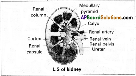
- Kidney is bean shaped structure, the outer surface of kidney is convex and inner surface is concave where it has a deep notch called hilum.
- A longitudinal sections of the human kidney shows two distinct regions namely the outer cortex and the inner medulla.
- Medulla is divided into multiple cone shaped masses of tissue called renal pyramids. The renal pyramids are separated by the projections of the cortex called columns of Berlin.
- The tips of the pyramids are renal papilla.
- Renal papilla projects into cup like calyces, formed by the funnel shaped pelvis, which continues out as the ureter. Ureter carries urine into urinary bladder.
- In cortex and medulla, nearly one million nephrons are present. They are structural and functional units of kidney. They are embedded in the loose connective tissue of cortex and medulla.
- In addition, kidney contains a network of blood capillaries, lymph sinuses and intestitial fluid in intra cellular spaces.
- The kidney gets blood supply through renal artery and blood from kidney is carried out by renal vein.
Question 5.
Explain micturition.
Answer:
The process of passing out of urine is called micro nutrition and the neural mechanism involved is called micturition reflex.
Urine is formed by the nephrons is ultimately carried to the urinary bladder where it is stored till a voluntary signal is given by the central nervous system (CNS). This sighal is initiated by the stretching of the urinary bladder as it gets filled with urine. In response, the stretch receptors on the walls of the bladder send signals to the CNS. The CNS passes on motor messages to initiate the contraction of smodth muscles of the bladder and simultaneous relaxation of the urethral sphincter, causing the release of urine.
Question 6.
What is the significance of juxta glomerular apparatus (JGA) in kidney function?
Answer:
Macula densa together with JG cells form juxtaglomerular apparatus (JAG). JAG plays a complex regulating role. A fall in glomerular blood flow or glomerular blood pressure or GFR can activate JG cells to release an enzyme called renin into the blood. This catalyses the conversion of angiotensinogen into angotensin-I which is further converted into angiotensin-II by angiotensin converting enzyme. Angiotensin-II, being a powerful vasoconstrictor increase the glomerular blood pressure and there by GFR.
Angiotensin-II also activates the adrenal cortex to release aldosterone. Aldosterone causes reabsorption of Na+ and water from distal convoluted tubule and collecting duct. To reduce loss through urine, and also promote secretion of K+ ions into distal convoluted tubule and collecting duct. It leads to increase in the blood pressure and GFR. This complex mechanism is generally known as renin – angiotensin- aldosterone system (RAAS).
Question 7.
Give a brief account of the counter current mechanism.
Answer:
Mammals have the ability to produce concentrated urine. The Henle’s loop and vasa recta plays an important role in this. The flow of the renal filterate in the two limbs of Henle’s loop is in opposite directions and thus form counter current. The flow of blood through vasa recta is also in counter current pattern. The proximity between the Henle’s loop and vasa recta, as well as the counter currents of renal fluid and blood in them help in maintaining an increasing osmolarity towards the inner medullary interstitium.
This gradient is mainly caused by NaCl and urea. NaCl passes out the ascending limb of Henle’s loop, and it enters the blood of the descending limb of vasa recta. NaCl is returned to the intestitium from the ascending portion of the vasa recta. Similarly small amounts of urea enter the thin segment of ascending limb of Henle’s loop which is transported back to the interstitium, from the collecting duct. Transport of these substances facilitated by the special arrangement of Henle’s loop and vasa recta.is called the counter current mechanism.
This mechanism helps to maintain a concentration gradient .in the medullary interstitium. Presehce of such interstitium gradient help easy passage of water from the collecting duct, there by concentrating the urine.
![]()
Question 8.
Explain the auto regulatory mechanism of GFR.
Answer:
Auto Regulation of GFR: *The kidneys have built in mechanisms for the regulation of glomerular filtration rate. One such efficient mechanism is carried out by juxta glomerular apparatus. Juxta glomerular apparatus is a special region formed by cellular modifications in the distal convoluted tubule and the afferent arteriole at the location of their contact.
A fall in GFR can activate the juxta glomerular cells to release an enzyme called renin, which catalyses the conversion of angiotensinogen into angiotensin-I and further converted to angiotensin-II by action of an enzyme angiotensin converting enzyme. Angiotensin-II’ stimulate the adrenal cortex to secrete aldosterone. Aldosterone causes reabsorption of Na+ and water from DCT and collecting duct to reduce loss through urine and also promotes the secretion of K+ ions into the DCT and CD (collecting duct). It leads.an increase in the blood pressure and GFR.
Question 9.
Describe the role of liver, lungs and skin in excretion.
Answer:
In addition to the kidneys, liver, lungs and skin also play an important role in the elimination of excretory wastes.
a) Liver :
Liver is the largest gland in our body, secretes bile, containing substances like bilirubin, biliverdin, cholesterol, degraded steroid hormones, vitamins and drags. Most of these substances ultimately pass out along with digestive wastes.
b) Lungs :
Lungs remove large amounts of C02 (18 litres 1 day), various, volatile materials and significant quantities of water.
c) Skin :
Human skin possesses two types of glands namely sweat and sebaceous glands for the elimination of certain substances through their secretions.
- Sweat produced by the sweat glands is a watery fluid containing NaCl, small amount of urea, lactic acid etc.,
- Sebaceous glhnds eliminate certain substances like sterols, hydrocarbons and waxes through sebum. This secretion provides a protective oily covering for the skin.
Question 10.
Name the following.
Answer:
a) A chordate animal having protonephridial type excretatory structures Cephalo chordate.
b) Cortical portions projecting between the medullary pyramids in the human kidney. Columns of Bertini
c) Capillary network paralleing the loop of Henle. Vasa recta.
d) A non chordate animals having green glands as excretory structures. Crustaceans.
Long Answer Questions
Question 1.
Describe the excretory system of man, giving the structure of a nephron.
Answer:
In humans, the excretory system consists of a pair of kidney, a pair of ureters, a urinary bladder and urethra.
Kidney :
Kidneys are reddish brown, bean shaped structures, situated on either side of the vertebral column between the levels of last thoracic and third lumbar vertebrae in a retroperitoneal position. The right kidney is slightly lower than the left one due to the presence of liver.
The outer surface of the kidney is convex and the inner surface is concave, where it has a deep notch called hilum, the point at which the renal artery and nerves enter and renal vein and ureter leave. Each kidney is surrounded by a tough, fibrous tissue, called renal capsule.
Ureter :
These are slender whitish tubes, which emerges from the pelvis of the kidney. The ureter rundown and open into the urinary bladder.
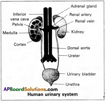
Urinary bladder :
Urinary bladder is a pear shaped like muscular organ. It tempirarily stores the urine, situated in the lower abdominal cavity. The neck of the bladder leads into the urethra. Urethra opens near the vaginal orifice in the female and through the penis in the male.
Structure of nephron:
Each kidney has nearly one million nephrons. These are structural and functional units of kidney, embedded in the loose connective tissue of cortex and medulla. Nephron consist of malpighian body and renal tubule.
I) Malphigian body :
It begins in the cortex of the kidney. It contains Bowman’s capsule and glomerulus.
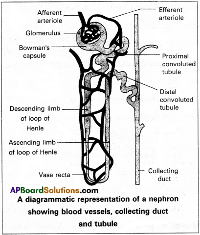
a) Bowman’s capsule :
It is a thin walled, double layered cup. The inner wall of the Bowman’s capsule has certain unique cells called podocytes.
b) Glomerulus :
It is a dense network of capillaries in the cup of Bowman’s capsule. Afferent arteriole of renal artery enter the cavity of Bowman’s capsule and split into five branches.
They unite and come out of the Bowman’s capsule as an afferent arteriole.
The podocytes of inner wall of Bowman’s capsule wrap around each capillary. The podocytes are arranged in an intricate manner so as to leave some minute spaces called filteration slits. The endothelium cells of capillaries have numerous pores called fenestrations.
II) Renal tubule:
It is narrow, delicate tubule arises from the posterior part of Bowman’s capsule known as neck. It opens into along narrow convoluted tubule with three parts like proximal convoluted tubule, Loop of Henle and Distal convoluted tubule.
a) Proximal convoluted tubule :
It is a lined by simple cuboidal epithelium with brush border to increase area of absorption.
b) Loop of Henle :
It is a hairpin like tubule present in medulla region. It consist of a descending limb and an ascending limb. The proximal part of the ascending limb is thin and the distal part is thick. The thick ascending limb continuous into the distal convoluted tubule.
c) Distal convoluted tubule (DCT) :
It is present in cortex. It is lined by simple cuboidal epithelium. The DCT continuous as the initial collecting duct in the cortex.
Collecting system :
Some initial collecting ducts unite to form straight collecting duct, which passes through the medullary pyramid. In the medulla, the tubes of each pyramid join and form duct of Bellini, which finally opens into tip of the renal papilla.
Capillary network of nephron :
The efferent arteriole emerging from the glomerulus forms a fine capillary network called the peritubular capillaries, around the renal tubule. The portion of the peritubular capillaries that surrounds the loop of Henle is called the vasa recta. The vasa recta is absent or highly reduced in the cortical nephrons. The juxta medullary nephrons possess well developed yasa recta.
![]()
Question 2.
Explain the physiology of urine formation.
Answer:
The formation of urine involves three main processes namely
- Glomerular Alteration
- Selective reabsorption
- Tubular secretion.
1) Glomerular Alteration :
It is Arst step in urine formation. The process of Alteration of blood, which occurs between glomerulus and lumen of the Bowman’s capsule due to difference in netpressure is called glomerular Alteration.
The hydrostatic pressure of blood while Aowing in the glomerulus is 60 mm Hg. It is opposed by glomerular colloidal osmotic pressure of 32 mm Hg and Bowman’s capsule hydrostatic pressure of 18 mm Hg.
The net filterate pressure is 10 mm Hg ( 60 – 32 + 18 = 10). This causes the Alteration of blood through the 3 layered filterate membrane formed by endothelium cells of glomerular capillary together with the basement membrane and podocytes of the Bowman’s cup. By the result of glomerular Alteration primary urine or renal Auid is collected in lumen of the Bowman’s capsule.
The primary urine contains almost all the constituents of plasma, except the proteins. The primary urine is hypotonic to the cortical Auid, it passes into the next part of renal tubule.
2) Selective reabsorption:
During the process of glomerular Alteration 125 ml/minute df primary urine is formed. Nearly 99% of which and essential substances are reabsorbed* by renal tubules called selective reabsorption. About 85% of filterate formed (primary urine) is reabsorbed in a constant unregulated manner called obligatory reabsorption.
3) Tubular Secretion :
During the formation of urine, the tubular cells secrete substances such as H+, K+ and NH3+ into the filterate. Tubular secretion is also an important step in the formation of urine as it helps in maintenance of ionic and acid base balance of the body fluids.
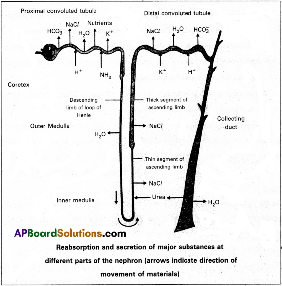
Mechanism of selective reabsorption and secretion takes place is different parts of nephrons.
a) In the proximal convoluted tubule :
Nearly all the essential nutrients and 70-80% of electrolytes and water are reabsorbed by this segment. Na+, glucose, amino acids, Cl– and other essential substances are reabsorbed into blood.
PCT also helps to maintain the pH and ionic balance of body fluids by selective secretion of H+ and NH3 into the filterate and by the absorption of HCO3– from it.
b) In the Henle’s loop :
Reabsorption in this segment is minmium.
- The descending loop of Henle is permeable to water and almost impermeable to electrolytes results the filterate concentration gradually increases.
- The ascending limb has two specialized regions, a proximal thin segment in which NaCl diffuses out into interstitial fluid passively, and distal thick segment, in which NaCl is actively pumped out.
The ascending limb is impermeable to water. Thus the filterate becomes progressively more dilute as it moves up to the cortex i.e., towards the DCT.
In the Distal convoluted tubule (DCT) :
It is permeable to water and ions. The reabsorption of water is variable depending on several conditions and is regulated by ADH. DCT is also capable of reabsorption of HCO3– and selective secretion of H+ and K+ ions and NH3+ into DCT from peritubular network, to maintain the pH and sodium – potassium balance in tHe blood.
In the collecting duct (CD) :
Considerable’amount of water could be reabsorbed from this region to produce concentrated urine. This segment allows passage of small amount of urea to medullary interstitium to keep up its osmolarity. It also plays a role in the maintenance of pH and ionic balance of blood by the selective secretion of H+ and K+ ions.
The renal fluid after the process of facultative reabsorption in the CD, influenced by ADH, constitute the urine, that is sent out. Urine in the CD is hypertonic to the plasma of blood.