AP Inter 2nd Year Zoology Study Material Chapter 8 Applied Biology

Andhra Pradesh BIEAP AP Inter 2nd Year Zoology Study Material 8th Lesson Applied Biology Textbook Questions and Answers.

AP Inter 2nd Year Zoology Study Material 8th Lesson Applied Biology

Very Short Answer Questions

Question 1.
What are the factors constitute dairying?
Answer:

  1. Selection of good breed having high yielding potential, combined with disease resistance ones.
  2. Proper housing with adequate water, feed, ventilation suitable temperature etc.

Question 2.
Mention any two advantages of inbreeding.
Answer:

  1. Inbreeding increases homozygosity. Thus inbreeding is necessary if we want to evolve a pure line animal.
  2. It helps in the accumulation of superior genes and elimination of less desirable genes.

Question 3.
Distinguish between out-cross and cross-breed.
Answer:
Out cross :
The offspring formed by mating of animals within the same breed, but having no ancestors on either side of pedigree for 4-6 generations.

A single out cross helps to overcome inbreeding depression.

Cross breed :
The offspring formed by a mating between superior males of one breed and superior females another breed.

Cross breed shows desirable qualities of two different breeds to be combined.

Question 4.
Define the terms layer and broiler.
Answer:
Layer :
The birds which are raised exclusively for the production of eggs are called layers.

Boiler :
The birds which are raised only for their meat are called broilers.

Question 5.
What is apiculture?
Answer:
Apiculture is the maintenance of hives of honeybees for the production of honey and wax.

Apiculture is an age-old cottage industry.

AP Inter 2nd Year Zoology Study Material Chapter 8 Applied Biology

Question 6.
Distinguish between a drone and worker in honey bee colony.
Answer:

Drones worker bees
1) These are fertile males. 1) These are sterile female.
2) These are developed from unfertilized ova by male parthenogenesis. 2) These are developed from fertilized eggs.
3) These are short lived. 3) These live for two and three months.

Question 7.
Define the term Fishery.
Answer:
Fishery is an industry devoted to the catching, processing for storage in freezers and selling of fish, shellfish or any other aquatic animals for human consumption.

Question 8.
Differentiate aquaculture and pisciculture.
Answer:

Aquaculture Pisciculture
Culturing of fishes and other aquatic organisms under regulated conditions to achieve better production. Culturing of exclusively fin fishes under regulated conditions to achieve better production.

Question 9.
Explain the term hypophysation.
Answer:
Making the fishds to breed artificially to meet the demand of carpseed as called hypophysation.

Question 10.
List out any two Indian carps and two exotic carps.
Answer:
Indian carps :

  1. Catla catla (catla)
  2. Cirrhinus mrigala (mrigal)

Exotic carps :

  1. Grass carp
  2. Silver carp

Question 11.
Mention any four fish by-products.
Answer:

  1. Shark and cod liver oils
  2. Fish guano
  3. Shagreen
  4. Isinglass.

Question 12.
How many aminoacids and polypeptide chains are present in insulin?
Answer:
Human insulin is made up of 51 aminoacids arranged in two polypeptides.
– polypeptide chain A with 21 aminoacids
– Polypeptide chain B with 30 aminoacids.

Which are held together by disulphide linkages.

Question 13.
Define the term vaccine.
Answer:
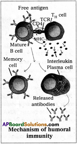
Vaccine is biological preparation that improves immunity to a particular disease. A vaccine typically contains live attenuated an inactivated disease causing organism. The toxins or one of the surface proteins of pathogens are also used in the preparation of vaccines.

Question 14.
Mention any two features of PCR.
Answer:

  • Very low concentration of bacteria or viruses can be detected by amplification of their nucleic acids by PCR.
  • PCR helps to detect very low amounts of DNA by amplification of the small DNA fragments.

PCR is now routinely used for detection of HIV in suspected cases, detection of mutations and genetic disorders.

Question 15.
What does ADA strand for? Deficiency of ADA causes which disease?
Answer:
ADA stands for adenosine deaminase. Deficiency of adenosine deaminase (ADA) causes severe combined immuno deficiency (SCID).

Question 16.
Define the term transgenic animal.
Answer:
Animals that have their own genome and had their DNA manipulated to possess and express an extra or foreign gene is known as transgenic animals.

Question 17.
What is popularly called “Guardian anger of Cell Genome?
Answer:
The protein p53 is a tumor suppressor protein, which plays an important role with reference to the ”G1 check point”. In the regulation of cell division cycle. It guards the integrity of the DNA. So it is also called guardian angel of cell’s genome.

Question 18.
List out any four features of cancer cells.
Answer:

  • Loss of contact inhibition
  • Reduced intra cellular adhesion
  • Immortalization
  • Loss of anchorage dependence

Question 19.
How do we obtain radiographs?
Answer:
A beam of X-rays is produced by an X-ray generator and is projected on the body parts. X-rays that pass through the body parts are recorded on a photographic film. Photographs developed using X-rays are known as radiographs.

AP Inter 2nd Year Zoology Study Material Chapter 8 Applied Biology

Question 20.
What is tomogram?
Answer:
Tomogram is a recorded image formed by computed tomography which shows the 3-D cross sectional pictures of the part of the body and displays the picture on the screen.

Question 21.
MRI scan is harmless. Justify.
Answer:
MRI does not use ionizing radiation, as involved in X-rays, and is generally safe and harmless procedure.

Question 22.
What is electrocardiography and what are the normal components of ECG?
Answer:
Electrocardiography is a commonly used, non invasive procedure for recording electrical changes in the heart.

Normal components of ECG:
(i) Waves (ii) Intervals (iii) Segments (iv) Complexes.

Question 23.
What does prolonged F-R interval indicate?
Answer:
Prolonged P-R interval indicates delay in conduction of impulses from S-A node to the A-V node.

P-R interval is prolonged in bradycardia.

Question 24.
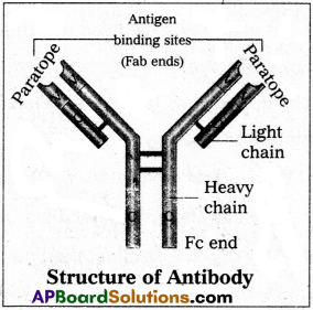
Differentiate between primary and secondary antibodies.
Answer:

Primary antibodies Secondary antibodies
1) These antibodies are formed against the specific antigen. 1) These antibodies are formed against the foreign primary antibody.
2) These antibodies reacts with the antigens of interest. 2) These antibodies react with the primary antibodies.

Question 25.
Which substances in a sample are detected by direct and indirect ELISA respectively.
Answer:

  1. Direct ELISA – used to detect antigens present in the sample.
  2. Indirect ELISA – used to detect antibodies present in the sample.

Short Answer Questions

Question 1.
What are the various methods employed in animal breeding to improve livestock?
Answer:
Animal breeding is the method of mating closely related individuals.
There are broadly two methods in animal breeding. (1) In breeding (2) Out breeding
1) In breeding:
When crossing is done between animals of the same breed it is called in breeding. In breeding is of two types (a) Close breeding (b) Line breeding.
a) Close breeding:
Close breeding is mating between male parent and female offspring and/or female with male offspring.

b) Line breeding :
Line breeding is the selective breeding of animals for a desired feature by mating them within a closely related line. It leads to upgrading of a desired commercial character.

2) Out breeding:
Out breeding is the breeding of the unrelated animals. Out breeding is of three types (a) Out-crossing (b) Cross-breeding (c) Interspecific hybridisation.

a) Out-crossing :
Mating of animals within the same breed, but having no common ancestors on either side of pedigree for 4-6 generations. The off spring of such mating is known as an out-cross.

b) Cross-breeding :
In this method, superior males of one breed are mated with superior females of another breed. The offspring of such a mating is said to be a cross breed.

c) Interspecific hybridisation :
In this method, male and female animals of two different related species are mated. The progeny may combine desirable features of both the parents and is different from both the parents.

Question 2.
Define the term breed. What are the objectives of animal breeding ?
Answr:
Breed:
A breed is a group of animals related by descent and similar in most characters such as general appearance, size, configuration and features with other members of the same species.

Jersery and Brown Swiss are example of foreign breeds of cattle. These two varieties of cattle have the ability to produce abundant quantities of milk. This milk is very nutritious with high protein content.

Objects of animal breeding :

  1. To produce disease resistant animals.
  2. Increase in the quality and quantity of milk, meat, wool etc.,
  3. Fast growth rate.
  4. Enhanced productive life by improving the genetic merit of livestock.
  5. Early maturity
  6. Economy of feed

Question 3.
Explain the role of animal husbandry in human welfare.
Answer:
Animal husbandary deals with the scientific management of livestock. It includes various aspects such as feeding, breeding and control diseases to raise the population of livestock. Animal husbandary usually includes buffaloes, cows, pigs, horses, cattle, sheep, camels, goats, poultry, fish etc which are useful for humans in various ways.

These animals are managed for production of commercially important products such as milk, meat, wool, egg, honey, silk etc. The increase in human population has increased the demand of these products. Hence it is necessary to improve the management of livestock scientifically. ,

Question 4.
List out the various steps involved in MOET.
Answer:
The following are the steps involved in Multiple Ovulation and Embryo Transfer /MOET):

  • A cow is administrated hormones, with FSH like activity.
  • This induces follicular maturation and super ovulation.
  • In Super ovulation instead of one egg, which they produce per cycle, they produce 6 – 8 eggs.
  • The cow is either mated with elite bull or artificially inseminated.
  • The embryos are at 8-32 called stages are recovered non-aurgically and transferred to surrogate mother, when the embryo develops into complete animal.

Now the genetic mother is ready for another round of super ovulation. This technology is in use for cattle, sheep, rabbits, buffaloes etc. to produce high yielding ones.

Question 5.
Write short notes on controlled breeding experiments.
Answer:
Controlled breeding experiments are carried out using artificial insemination and multiple ovulation and embryo transfer technology.

  • In this technique the semen is collected from superior bulls. This semen can be used immediately or can be frozen and used later period. It can be transported in a frozen form to place where a female is housed.
  • Meanwhile a cow or animal is administered hormones, with FSH like activity.
  • These hormones induces follicular maturation and super ovulation.
  • Now the cow is artificially inseminated for fertilisation.
  • The embryos are at 8-32 celled stages are recovered non-surgically and transferred to surrogate mother uterus for further development.

This technology is use for cattle, sheep, rabbits, buffaloes etc. By using this method we can produce high milk and meat yielding animals and also control the venereal diseases.

AP Inter 2nd Year Zoology Study Material Chapter 8 Applied Biology

Question 6.
Explain the important components of poultry management.
Answer:
Important components of poultry management:

Selection of disease free and suitable breeds:
The selected’breeds should be disease free and get acclimatised to a wide range of climatic conditions. Eg: In India Hybrid layers-BV 300, Hyline, Poona – Pearls etc., Broiler strains – Hubbard, Vencobb etc.

Feed management:
Balanced diet is must to maximise the yield. Brooder, chick mash, grower mash, prelayer mash and layer mash are fed to layers at different stages. Likewise pre starter mash, starter mash and finish mash are the feed given to broilers. Safewater should be supplied through waterers at all times.

Health care :
Vaccination against viral diseases and using antibodies for both bacterial and fungal diseases.

In addition to the above hygiene, proper and safe farm conditions ensure better produce.

Question 7.
Discuss in brief about ‘AvianFlu’.
Answer:
AvianFlu or birdFlu is an important disease affecting poultry birds and man.

Causative organism :
AvianFlu or birdFlu is caused by an “avianFlu virus” the H5NI. The virus that causes the bird infection infects humans too. It is a pandemic disease.

Mode of infection:
Infection may be spread simply by touching contaminated surfaces. Birds infected by this type of influenza, continue to release the virus as in their faeces and saliva for as long as 10 days.

Symptoms:
In humans it causes typical-flu-like symptoms, include cough, diarrhoea difficulty in breathing, fever, headache, malaise, muscle aches and sore throat.

Prevention :

  • Avoiding consumption of under cooked chicken.
  • People who work for poultry birds should use protective clothing and special breathing masks.
  • Complete culling of infected flock by burying or burning them.

Question 8.
Explain in brief about queen bee.
Answer:

  • Queen bee is the largest individual in the colony.
  • It is a fertile diploid female, one per bee hive and the egg layer of the colony.
  • She lives for about five years and her only function is to lay eggs.
  • The queen bee during its nuptial flight receives sperms from a drone and stores in the spermathecae and lays two types of eggs, the fertilised and unfertilised.
  • All fertilised eggs develop into females.
  • All the larvae developing from the fertilised eggs are fed with the royal jelly for first four days only. Afterwards royal jelly is fed only to the bee that is bound to develop into next queen, whereas the other larvae fed on bee bread become workers.

Question 9.
Honey bees are economically important – justify.
Answer:

  • Honeybees are economically important insects in the world. Because honeybee products like Honey, wax, propolis and beevenom have more economic importance.
  • Honey – It is a rich source of fructose, glucose, water minerals and vitamins.
  • Bee’s wax – It is used in the preparation of cosmetics, polishes of various kinds and candles.
  • Propolis – Propolis is used in the treatment of inflammation and superficial bums.
  • Bee’s Venom – Which extracted .from the string of worker bees is used in the treatment of rheumatoid arthritis.
  • Pollination – Bees are the pollinators of our crop plants such as sunflower, Brassica, Apple and Pear.

Question 10.
What are the various factors required for Bee keeping?
Answer:
Bee keeping or apiculture is the maintenance of hives of honeybees for the production of honey and wax.

Factors required for successful Bee keeping :

  1. Knowledge of nature and habits of honeybees.
  2. Selection of suitable location for keeping the beehives. ‘
  3. Raising a hive with the help of a queen and small group of worker bees.
  4. Management of beehives during different seasons.
  5. Knowledge of handling procedures and collection of honey and bee wax.

Question 11.
Fisheries have carved a niche in Indian economy. Explain.
Answer:
Fisheries have carved a niche in Indian economy, as fisheries have more economic importance.

As food :
Fish meat, in general is a good source of proteins, vitamins, minerals and rich in iodine. Tunas, shrimps and crabs are not only edible but also have export value.

Byproducts :

  1. Shark and Cod liver oils – are good source of vitamins A and D.
    Oils from Sardine and Salmon- are good source of Omega 3 – fatty acids.
  2. Fish guano from Scarp fish – used as fertilizer.
  3. Shagree and I$inglass – used in clarification of wines.

Question 12.
Explain in brief structure of Insulin.
Answer:
AP Inter 2nd Year Zoology Study Material Chapter 8 Applied Biology 1
Insulin is a poly peptide hormone produced by the β – cells of islets of langerhans of pancreas. It is the first protein produced by recombinant DNA technology.

Structure of Insulin :
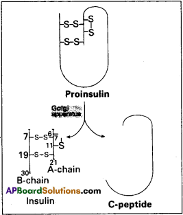
Human insulin is made up of 51 aminoacids arranged in two polypeptide chains. The chain A has 21 aminoacids while chain B has 30 aminoacids. Both are held together by two interchain disulfide bridges, connecting A7 to B7 arid A20 to B19. In addition, there is an intrachain disulfide link in chain A between the aminoacids 6 and 11.

In mammals, including humans, insulin is synthesized as a pro-hormone, which contains an extra stretch called the ‘c’ peptide. This ‘c’ peptide is not present in the mature insulin and is removed during maturation into insulin. .

Question 13.
Define Vaccine and discuss about types of Vaccines.
Answer:
A Vaccine is a biological preparation that improves immunity to a particular diseases. A Vaccine typically contains inactive or attenuated disease causing microorganisms. The toxin or one of the surface proteins of the microorganisms are also used in preparing vaccines.

Types of Vaccines :
1) Attenuated whole agent vaccines :
They contain disabled line microorganisms. Mostly they are antiviral. Eg: Vaccines against Yellow fever, measles, rubella and mumps and the bacterial disease such as typhoid.

2) Inactivated whole agent vaccines :
They contain killed microbes. Eg : Vaccines against influenza, cholera, hepatitis A, rabies etc.

3) Toxoids:
They contain toxoids which are inactivated exotoxins of certain microbes.
Eg : The vaccines against diphtheria and tetanus.

Question 14.
Write in brief the types of gene therapy. .
Answer:
Gene therapy is the insertion of genes into an individual’s cells and tissues to treat a
There are two approaches to achieve gene therapy :

  1. Somatic line therapy
  2. Germ line gene therapy

1) Somatic line therapy:
In this type of therapy, functional genes are introduced into somatic cells of a patient. The approach is to correct a disease phenotype by treating defect in somatic cells in the affected person. The changes effected in this type of gene therapy are nonfinheritable.
Somatic line therapy is of two types :
a) Ex-vivo gene therapy:
In which the cell are collected from patient, modified outside the body and then transplanted back Eg: SCID.

b) In-vivo gene therapy :
In this therapy, the genes are changed in cells, while they are still inside the body Eg : Cystic fibrosis.’

2) Germ line gene therapy:
In this type of therapy, functional genes are introduced into sperms or ova and are thus integrated into their genomes. Therefore the changes or modifications become heritable. Due to various technical and ethical reasons, the germ line gene therapy remained at infant stage.

Question 15.
List out any four salient features of cancer cells.
Answer:
Salient features of Cancer cells :
Loss of contact inhibition :
Normal cells in a culture stop growing when their plasma membranes come into contact with one another. This inhibition of growth after contact is called contact inhibition. Cancer cells lose this property.

Reduced intracellular adhesion :
When normal cells growing in medium, the cells are joined by intracellular adhesion proteins called cadherins. They are missing in Cancer cells.

Immortalisation :
Normal cell culture does not survive indefinitely. They undergo apoptosis. Where as Cancer cells do not undergo apoptosis.

Loss of anchorage dependence :
Most normal cells must be attached to a rigid substratum in order to grow Cancer cells can grow even when they are not attached to the substratum.

Increased growth of blood vessels :
When tumors grow in size diffusion of oxygen and nutrients become restricted and so tumors resort to attracting more blood vessels from their surrounding matrix.

AP Inter 2nd Year Zoology Study Material Chapter 8 Applied Biology

Question 16.
Explain the different types of cancers.
Answer:
Based on the origin Cancers are classified into :
1) Carcinomas :
These are malignant tumor of epithelial cells. They are originating from the epithelial tissues of skin, lining of the respiratory, digestive, urinary and genejal systems or cells from various glands breast and nervous tissue etc. 85% of Cancers are Carcinomas.

2) Sarcomas :
These are malignant tumors of connective tissues or organs that originate from mesoderm. About 2% of tumors are Sarcomas.

3) Leukemias :
These are malignant tumors of stem ceils of hematopoietic tissues, resulting in unrestrained production of WBC. They are liquid tumors. About 4% of Cancers are Leukemias.

4) Lymphomas :
These are malignant tumors of secondary lymphoid organs like spleen, and lymphnodes. About 4% of Cancers are Lymphomas.

Question 17.
Write about the procedure involved in MRI. X jmsin
Answer:
MRI Scan is a diagnostic radiology technique that uses magnetism, radiowaves and a computer to produce images of body components.

Procedure :
MRI Scanner is giant circular magnetic tube.

  • The patient is placed on a movable bed that is inserted into the magnet.
  • Human body is mainly composed of water which contains two protons.
  • The magnet creates a strong magnetic field that makes these proton align with the direction of the magnetic field.
  • A second radiofrequency electromagnetic field is then turned on for a brief period. The protons absorb some energy from these radio waves.
  • When this second radio frequency emitting field is turned off, the protons release energy at a radio frequency which can be detected by the MRI scanner.
  • Different types of tissues emit different quanta of energy. Abnormal tissues such as tumors can be detected because the protons in different types of tissues return to their equilibrium state at different rates.
  • Tissues of bone with less water content look different in MRI, and pathological tissues also can be detected.

The information received is processed by computer and generated an image.

Question 18.
Write briefly about different waves and intervals in an ECG. X
Answer:
ECG (electrocardiography) is commonly used, non-invasive procedure fro recording electrical changes in the heart.

The graphic record which is called an electrocardiogram, shows the series of waves that occur during each cardiac cycle.

The normal ECG consists of (i) Waves (ii) Intervals (iii) Segments (iv) Complexes.

i) Waves :

  • The waves in a normal record are named P, Q, R, S and T in that order.
  • A typical ECG tracing of a normal heartbeat consists of (I) a ’P’ wove (II) a ‘QRS complex of waves’ (III) a T Wave.
  • P wave: It represents the atrial systole and shows that the impulse is passing through atria. The duration of P. Wave is 0.1 sec.
  • QRS complex of wave : It represents ventricular systole. Q wave is small negative., R-wave is tall positive and S wave is a negative wave. Its duration is 0.08 to 0.1 sec.
  • T wave: It represents the ventricular repolarization. It is a positive wave,’its duration is 0.2 sec.

ii) Intervals:
P-R intervals :
P-R intervals is the interval between the onset of p wave and the onset of Q wave. P-R interval is normally. 0.12 – 02 sec.

Q-T intervals :
The interval between the onset of Q wave and the end of the T-wave. It represents the electrical activity in muscle of the ventricles. It lasts for about 0.4 seconds.

R-R intervals:
It signifies the duration of one cardiac cycle and lasts for about 0.8 sec

Segments :
S-T segment is the time period between the end of the ‘S’ wave and the onset of the T-wave. It is an isoelectric or zero voltage period.

Question 19.
Discuss briefly the process of indirect ELISA.
Answer:
Enzyme linked immunosorbent assay is a tool of clinical immunology to detect, antigens or antibodies in a given sample. ELISA is of two types (1) Direct ELISA (2) Indirect EUSA.

Indirect ELISA:
It is used to detect antibodies present in the serum of the patient or given sample.

Protocol

  • A known antigen is added to the well, which absorbed on the surface of well.
  • Patients antiserum is added to AG coated well.
  • Allowed to react antibodies present in the serum with the antigen, coated on the surface of the well.
  • Washed the well to remove the any unbound free antibodies present in the well.
  • Enzyme linked antihuman serum globulins are added. They bind to the antibody which is already bound to the antigen.
  • Washed it to remove excess antibodies present m the well.
  • Enzyme substrate is added and the reaction produces a visible colour change which can be measured by a spectro photometer.

If there are no antibodies (i.e., anti HIV antibodyies in the serum sample, there is no binding of primary antibodies to the antigens and so enzyme linked secondary antibodies do not bind to the primary antibodies. There cannot be any enzymatic reaction and so no colour change is observed the test is said to be negative.

Question 20.
Write short note on EEG.
Answer:
Electro encephalography is the process of recording the electrical activity of the brain with help of an EEG machine and some electrodes placed all over the scalp.

The waves recorded by an EEG consist of synchronized waves which are common in normal healthy people and, in certain neurological conditions the waves are desynchronized. The wave pattern can be broadly classified into alpha, beta, delta and theta wave pattern.

Alpha waves :
They are rhythmical 8-13 cycleslsec. This type of Wave pattern is seen in persons who are drowsy or sleepy with closed eyes.

Beta waves:
These waves occur at a high frequency of 13-40 cycleslsec their amplitude is low. There are desynchronized waves recorded in person who are mentally very active and tense.

Delta waves :
Their frequency is quite low i.e., less than 3 cycleslsec. They have high amplitude. They are common in early childhood in awaken condition. In adults, they occur in deep sleep, epilepsy, mental depression etc. .

Theta waves:
Their frequency is between 4 and 7 cycleslsec. These waves are common in children of less than 5 years of age and emotional stress in adults.

Uses :

  • EEG is useful tool in diagnosing neurological apd sleep disorders.
  • The diagnostic application of EEG is the diagnosis of epilepsy.
  • EEG is also useful in the diagnosis of coma and brain death.

Long Answer Questions

Question 1.
Write in detail about outbreeding.
Answer:
Out breeding is the breeding of the unrelated animals, it is the cross between different breeds.
Out breeding is of three types

  1. Out crossing
  2. Cross breeding
  3. Inter specific hybridisation.

1) Out crossing :
It is the practice of mating of animals with in the same breed, but having no common ancestors on either side of the pedigree for 4-6 generations. The offspring of such a mating is known as an outcroas. It is the best breading method for animals that are below average in milk production, growth rate etc.

2) Cross breeding :
In this method, superior males of one breed are mated with superior females of another breed. The offspring of such a mating is said to be a cross breed. Cross breeding allows the desirable qualities of two different breeds to be combined. The progeny is not only used for commercial production but also inbreeding and selection to develop stable breeds which may be superior to existing breeds.
Eg : Hisardale is a new breed of sheep developed by crossing Bikaneri ewes and Marino rams. ‘ . .

3) Inter specific hybridisation :
In this method, male and female animals of two different related species are mated. The progeny may combine desirable features of both the parents and is different from both the parents.
Eg: 1) When a male donkey is crossed with a female horse, it leads to the production of “mule” (sterile/
2) When a male horse is crossed with a female donkey “hinny” (sterile) is produced. Mules have considerable economic value.

AP Inter 2nd Year Zoology Study Material Chapter 8 Applied Biology

Question 2.
Explain in detail clinical inferences from ECG. –
Answer:
ECG is commonly used, non-invassive procedure for recording electrical changes in the heart The graphic record is called an electrocardiogram, shows the series of waves that occur during each cardiac cycle.

Normal ECG consist of waves, intervals, segments and complexes.

Waves :
A typical ECG tracing normal heart beat consist of a ‘P’ wave a QRS complex of waves, a T wave.

P wave :
It represents the atrial systole and shows that the impulse is passing through atria. The duration of P wave is 0./ sec.

QRS complex of wave:
It represents ventricular systole Q wave is small negative, R-Wave is tall positive and S-yvave is a negative wave. It’s donation is 0.08 to 0.1 sec.

T wave :
It represents the ventricular repolarization. It is a positive wave, its duration is 0.2 sec.

Intervals:
P – R intervals :
It is the interval between 9nset of P wave and onset of Q wave. P-R interval is normally 0.12-0.2 sec.

Q – T intervals :
The interval between the onset of Q wave and the end of the • T-wave. It represents the electrical activity in muscle of the ventricles. It lasts for about 0.4 sec.

R – R intervals :
It signifies the duration of one cardiac cycle and lasts for about 0.8 sec . .

Segments :
S-T segment is the time-period between the end of the ‘S’ wave and the onset of the T-wave. it is an isoelectric or zero voltage period.

Clinical inferences from ECG :

  1. Enlarged P wave – indicates enlarged atria
  2. Variation in the duration, amplitude and morphology of the QRS complex – indicates disorders such as block of conduction of impulses through the branches of the bundle of His.
  3. Prolonged P-R interval duration – indicates delay in conduction of impulses from S-A node to the A – V node.
    P-R interval is prolonged in bradycardia and shortened in tachycardia.
  4. Prolonged Q-T interval – indicates myocardial infraction and hypothyroidism.
  5. Shortened Q.T interval – indicates hyper calcemio.
  6. Elevated S – T segment – indicates myocardial infarction.
  7. Tall T wave – indicates hyperkalemia.
  8. Small, flat or inverted T wave – indicates hypokalemia.

AP Inter 2nd Year Zoology Study Material Chapter 7 Organic Evolution

Andhra Pradesh BIEAP AP Inter 2nd Year Zoology Study Material 7th Lesson Organic Evolution Textbook Questions and Answers.

AP Inter 2nd Year Zoology Study Material 7th Lesson Organic Evolution

Very Short Answer Questions

Question 1.
What is panspermia?
Answer:
According to Cosomozoic theory, life might have existed all over the universe in the form of resistant spores called panspermia. They might have reach the earth accidentally.

Question 2.
Define prebiotic soap. Who coined the term?
Answer:
Prebiotic soup word is coined by J.B.S.Haldane. Haldane called ocean as prebiotic soup. All the reactions which lead to the formation of organic molecules like sugars, amino acids, fatty acids purines and pyramidins etc., occurred in the ocean so it was described as the prebiotic soup.

Question 3.
How did-eukaryotes evolve?
Answer:
Eukaryotes evolved probably by two processes.

  1. Prokaryotes lived in the ancestral eukaryotes symbiotically and evolved into organelles such as mitrochondria and plastids.
  2. The endo membrane system of eukaryotes might have evolved by the infolding of plasma membrane of the ancestral prokaryotes.

Question 4.
What are the components of the mixture used by Urey and Miller in their experiments to simulate the primitive atmosphere?
Answer:
Urey and Miller -used a mixture of water vapour, methane, ammonia and hydrogen in their experiments to simulate the primitive atmosphere.

Question 5.
Mention the names of any four connecting links that you have studied.
Answer:
Connecting links clearly explain the path of evolution.

  • Peripatas between annelida and arthropoda.
  • Prototherians between reptilia and mammalia.

AP Inter 2nd Year Zoology Study Material Chapter 7 Organic Evolution

Question 6.
Define Biogenetic law, giving an example.
Answer:
It was proposed by Ernst Haeckel. It states that ontogeny repeats phylogeny which means the development history of an organism repeats the evolutionary history of its ancestor Eg : Tadpole larva of frog, it resembles fish both externally and internally. It possesses a, tail, gills and two chambered heart like that of fish. Later it metamorphoses into adult frog.

Question 7.
Define atavism with an example.
Answer:
Sudden appearance of some vestigial organs in a better developed condition is called atavism.
Eg : Tailed human baby

Question 8.
Cite two examples to disprove Lamarck’s inheritance of acquired characters.
Answer:

  1. Well developed muscles of athletes are not inherited to their children.
  2. Making perforations to pinna for wearing ornaments has been in practice in India for past several centuries. However no girl child is bom with readymade perforations . in their pinna.

Question 9.
Who influenced Darwin much in formulating the idea of Natural Selection.
Answer:
Three scientists influenced Darwin, they are :

  1. T.R.Malthus – On the principles of population
  2. Sir Charles lyell – Principle$ of Geology
  3. Alfred Russel Wallace – On the tendency of varieties do depart from original types.

Question 10.
What is common between Darwinism and Lamarckism?
Answer:
Presence of variations is common to Darwinism and Lamarckism.

Question 11.
What is meant by genetic load. Give an example.
Answer:
The existence of deletorious genes within the populations is called genetic load.
Eg: Genes for Sickle cell anaemia – The individuals homozygous for Sickle cell gene usually die early due to anaemia. .

Question 12.
Distinguish between allopatric and sympatric speciations.
Answer:
Allopatric speciation :
Speciation occurring in which first geographical isolation occurs, then secondly reproductive isolation occurs.

Sympatric speciation :
Reproduction isolation occurs without geographical isolation.

AP Inter 2nd Year Zoology Study Material Chapter 7 Organic Evolution

Question 13.
Mention the scientific names of ape like and man like earlier primates. Which man like primate first used hides to cover the bodies?
Answer:

  • Scientific name of ape like primate – Dryopithecus
  • Scientific name of man like primate – Ramapithecus
  • Homo neanderthalensis – They used hides to protect their body.

Short Answer Questions

Question 1.
Distinguish between homologous and analogous organs.
Answer:
1. Homologous organs :
The organs which have similar structure and origin but not necessarily the same function are called homologous organs. Eg : The appendages of vertebrates such as the flippers of whale, wings of bat, forelimbs of horse, paw of cat and hands of man have a common pattern in the arrangement of bones even though their external form and functions may vary to suit their mode of life.
AP Inter 2nd Year Zoology Study Material Chapter 7 Organic Evolution 1

2. Analogous organs :
The organs which have dissimilar structure and origin but perform the same function are called the analogous organs. Eg : Wings of butterfly and wings of a bird.
AP Inter 2nd Year Zoology Study Material Chapter 7 Organic Evolution 2

Question 2.
Write a short note on the theory of mutations.
Answer:
Theory of mutation was proposed by Hugo de Varies, who coined the term mutation.

Mutations are sudden, random inheritable changes that occur in organisms. Hugo de Varies observed this phenomenon in the evening primrose plant Oenothera lamarckiana, which shows different forms like

  • O. brevistylis – with small style.
  • O. levifolia – with smooth leaves.
  • O. gigas – with the giant form.
  • O. nanella – with dwarf form.

These characters are inherited to the progeny.

  • Darwin called mutations as sports of nature or saltations.
  • Bateson called them as discontinuous variations

Salient features of mutation theory :

  • Mutations occur from time to time in naturally breeding population.
  • Mutants differ from their parents.
  • Mutations are inheritable.
  • Mutations occur in any direction i.e., they are random.
  • They are discontinuous and not accumulated over generations.
  • They are full-fledged and so there are no intermediate forms.
  • They are subjected to natural selection.

Question 3.
Explain Darwin’s theory of natural selection with industrial melanism as an experimental proof.
Answer:
Darwin’s theory of natural selection does not explain what exactly evolution is, but explains how evolution might have occurred in nature. A classical example for natural selection is industrial melanism, exhibited by peppered moth-Biston betularia. These moths were available in two colours grey and black. Grey moths were abundant before industrial revolution in all over England. The reason for the existence of large number of grey moths during that period was camouflage on the trunks of trees.

But after the establishment of industries in England, black coloured moths were more and grey forms were less. This is due to pollution from industries in the form of soot turned barks of trees into black. So grey moths were easily identified and were more predated by birds. Thus grey moths decreased in number, black moths increased in the population.

Thus natural selection favoured the melanic moths (black) to reproduce more successfully. Natural selection of darker forms in response to industrial pollution is known as industrial melanism.

AP Inter 2nd Year Zoology Study Material Chapter 7 Organic Evolution

Question 4.
Discuss the role of different patterns of selections in evolution.
Answer:
Natural selection is a process by which the organisms are physically, physiologically and behaviorally better adopted to environment, survive and reproduce.

It is mainly of three types :

  1. Stabilizing selection,
  2. Directional selection,
  3. Disruptive Selection.

1. Stabilizing selection (Centrifetal selection) :
This selection operates in a stable environment. In this process, the organisms with average phenotype are preserved where as the extreme individuals from both the ends are eliminated. Hence it does not promote any evolutionary change that leads to specification, but maintains the phenotypic stability within the population over generations.
Eg : In England weight of newborn babies were studied in a large sample. Greater mortality was found in the babies whose weight was greater or lesser than the average weight of 81bs.

2. Directional selection :
This selection operates in an environment which gradually undergoes changes. It works by constantly removing the individuals from one end and constantly shifting the average value of fitness towards the other end of phenotypic distribution.
Eg : In case of giraffes the average value of the length of neck shifted towards the long-neck. Stabilizing selection takes over the directional selection. Once the average value of the phenotype coincides with the new optimum environmental conditions.

3. Disruptive selection (Centrifugal selection) :
This selection operates when homogenous environment changes into a heterogenous type. In this process, the organism of both the extreme phenotypes are selected while the individuals with average phenotype are eliminated. This can split the population into two or more sub-populations or species populations.
Eg : In California the sunflower population was divided into two sub-populations. One was adapted to dry area and the other was adapted to wet area.

Question 5.
Write a short note on Neo-Darwinism.
Answer:
It was proposed by Fischer, Sewall Wright, Mayr. According to this theory, five basic factors are involved in the process of Organic evolution. They are :

  1. Gene mutations
  2. Chromosomal mutations
  3. Genetic recombinations
  4. Reproductive isolation

1. Gene mutations :
Heritable changes in the structure of a gene are called gene mutations or point mutations. They alter the phenotypic character of the individuals. Thus, gene mutations tend to produce Variations in the offspring.

2. Chromosomal mutations:
Heritable changes in the structure of chromosomes called chromosomal mutations. They also bring about variations in the phenotype of organism which lead to the occurrence of variations in the offspring.

3. Genetic recombinations :
Recombinations of genes due to crossing over during meiosis are responsible for bringing genetic variability among the individuals of the same species.

4. Natural selection :
Natural selection does not produce any genetic changes, but it favours some genetic change while rejecting others.

5. Reproductive isolation:
The absence of gene exchange between population is called the reproductive isolation. It plays a great role in giving rise to new species and preserving the species integrity.

Question 6.
In a population of 100 rabbits which is in Hardy-Weinberg equilibrium, 24 are homozygous long eared. Short ears are recessive to long ears. There are only two alleles for this gene. Find out the frequency of recessive allele in the population.
Answer:
Number of rabbits in the population with Hardy-Weinberg equilibrium = 100
Number of dominant homozygous long eared rabbit = 24
Frequency of homozygous dominant long eared rabbits (P²) = [latex]\frac{1}{100}[/latex] × 24 = 0.24
Frequency of dominent allele (P) = 0.49
Frequency of recessive allele = q = 1 – 0.49 = 0.51

AP Inter 2nd Year Zoology Study Material Chapter 7 Organic Evolution

Question 7.
What is meant by genetic drift ? Explain genetic drift citing the example of Founder Effect.
Answer:
The change in the frequency of a gene that occurs merely by chance and not by selection, in small proportion is called genetic drift.

Suppose, for a gene with two alleles, the frequency of a particular allele is 1% (e = 0.01) the probability of losing that allele by chance from small population is more. The end result is either fixation or loss of that allele.

Genetic drift tend to reduce the amount of genetic variation within the population mainly by removing the alleles with low frequencies. It can examplified by Founder and Bottleneck effect.

Founder effect:
If a small group of individuals fro,m a population starts a new colony in an isolated region, those individuals are called the founders of the new population. The allelic frequency of their descendants are similar those of the founders rather than to their ancestral parent population.
Eg : O+ve blood group is present in nearly 100% of the red Indians. It means the forefathers of the Red Indians tribe were predominantly O+ve and they isolated themselves reproductively from other population.

AP Inter 2nd Year Zoology Study Material Chapter 6 Genetics

Andhra Pradesh BIEAP AP Inter 2nd Year Zoology Study Material 6th Lesson Genetics Textbook Questions and Answers.

AP Inter 2nd Year Zoology Study Material 6th Lesson Genetics

Very Short Answer Questions

Question 1.
What is Pleiotropy?
Answer:
The ability of a gene to have multiple phenotypic effects because it influences a number of characters simultaneously is known as Pleiotropy.
Eg: Phenylketonuria.

Question 2.
What are the antigens causing ‘ABO’ blood grouping? Where are they present?
Answer:
Isoagglutinogen A (antigen A) and Isoagglutinogen B (antigen B) are the antigens responsible for ABO blood grouping. These antigens are present on the surface of red blood cells.

Question 3.
What are the antibodies of ABO blood grouping? Where are they present?
Answer:
Isoagglutinin A (anti A) and Isoagglutinin B (anti B) are the antibodies of ABO blood grouping. These antibodies are present in the blood plasma.

Question 4.
What are multiple alleles?
Answer:
If a gene has more than two alleles then they are said to be multiple alleles.
Eg : In humans ABO blood groups are the best example for multiple allelism.

AP Inter 2nd Year Zoology Study Material Chapter 6 Genetics

Question 5.
What is erythroblastosis foetalis?
Answer:
Erythroblastosis foetalsis is an alloimmune condition that develops in an Rh positive foetus whose father is Rh positive and mother is Rh negative.

In this disorder the antibodies developed against the Rh antigen in mother, cross, placenta and destroy the RBC cells of the Rh+ve foetus during second pregnancy.

Question 6.
A child has blood group ‘O’. If the father has blood group A and mother blood group B, work out the genotypes of the parents and possible genotypes of the other off spring.
Answer:
Child blood group is ‘O’, and ‘O’ has the genotype I°I°. Hence, if father has blood group A and mother has blood group B, then the possible genotype of the parents will be IAI° and IBI° respectively.
AP Inter 2nd Year Zoology Study Material Chapter 6 Genetics 1

Genotypes of the off springs
IAIB – AB blood group
IAI° – A blood group
IBI° – B blood group
I°I° – O blood group

Question 7.
What is the genetic basis of blood types in ABO system in man?
Answer:
Three alleles of gene I are responsible for ABO blood grouping. They are IA, IB and I°.
IAIA / IAI° – for A blood group
IBIB / IB I° – for B blood group
IAIB – for AB blood group
I° I° – for O blood group

Question 8.
What is polygenic inheritance?
Answer:
Polygenic inheritance is a cumulative effect of two or more genes on a single phenotypic character. Eg: Skin colour in humans.

Question 9.
Compare the importance of Y-chromosome in human being and Drosophila.
Answer:
In human beings Y-chromosomes are responsible for the development of maleness.

In Drosophila Y-chromosome, lacks male determing factors, but contains only genetic information essential to male fertility.

Question 10.
Distinguish between heterogametic and homogametic sex determination systems.
Answer:

Heterogametic Homogametic
1. It is the condition in which two types of gametes are formed.
Eg: XY-in humans.
1. It is the condition, in which similar type of gametes are formed.
Eg: XX in females.
2. They play a very important role in deciding the sex of the off spring 2. It self, it can’t decide the sex of the progeny.

Question 11.
What is haplo-diploidy?
Answer:
It is a mechanism of sex determination. In this system the sex of the offspring is determined by the number of sets of chromosomes. Eg : Honeybees.

In honeybees fertilised eggs developed into female and unfertilised eggs developed into male. This means male have half the number of chromosomes ie., haploid and the females have double the number i.e., diploid hence the name haplo.-diploidy.

Question 12.
What are barr bodies?
Answer:
Barr bodies are condensed heterochromatin in one of the ‘X’ chromosome found in the somatic cells of diploid females. These were observed by Murray. L.Barr in female cats and Moor? and Barr in female human beings.

AP Inter 2nd Year Zoology Study Material Chapter 6 Genetics

Question 13.
What is Klinefelter’s syndrome?
Answer:
Klinefelter’s syndrome is caused by trisome in 23rd pair. A klinefelter’s male possesses an additional X chromosome along with the normal XY (i.e.,47 chromosomes).

Symptoms :
Hypogonadism, sterility, enlargement of breast, high pitched voice etc., Somatic cells of Klinefelter male exhibits barr bodies in their nuclei.

Question 14.
What is Turner’s syndrome?
Answer:
It is an allosomal disorder. The Karyotype is 45, it is due to monosomy in 23rd pair. These females have 42 autosomes and one X-chromosome.

Symptoms:
Short structure, gonadal dysgenesis, webbed neck, broad shield like chest and widely spaced nipples etc. Turners female does not show barr bodies.

Question 15.
What is Down syndrome?
Answer:
Down Syndrome is a genetic condition that causes delay in physical and intellectual development. The cause of this genetic disorder is the presence of an additional copy of the chromosome numbered 21.

Symptoms :
The affected individual is short, with small round head, furrowed tongue and partially open mouth. Physical and mental development is retarded.

Question 16.
What is Lyonisation?
Answer:
Lyonisation is a process by which one of two copies of X-ehromosome present in the body cells of female mammals is inactivated. The inactive X-chromosome is transcriptionally inactive called heterochromatic body.

Question 17.
What is sex-linked inheritance?
Answer:
The inheritance of a trait that is determined by a gene located on one of the sex chromosome is called sex linked inheritance.
Eg: Colour blindness, Haemophilia etc.

Question 18.
Define hemizygous condition?
Answer:
The condition in which thd genes are present on non-homologous portion of either X- chromosome (or) Y-chromosomes. For these genes, related alleles are absent on corresponding paired chromosomes.
Eg : X-linked genes and Y-linked genes in males.

Question 19.
What is crisscross inheritance?
Answer:
Crisscross inheritance is also called as skip generation inheritance. The X-linked recessive characters do not occur in one generation. They skip it off’in that generation and are expressed in the next generation. Eg: X-linked recessive characters – Colour blindness.

Colour blindness is transmitted from grandfather to his grandson through a carrier daughter.

AP Inter 2nd Year Zoology Study Material Chapter 6 Genetics

Question 20.
Why are sex – linked recessive characters more in male human beings?
Answer:
Sex linked recessive characters are more in males because these genes located in the X-chromosome. Male possess only one X-chromosome and female possess two ‘X’ chromosomes. So male needs only one copy of the mutant allele to express the phenotype.

Question 21.
Why are sex – linked dominant characters more in female human beings?
Answer:
In sex-linked dominant inheritance, the gene responsible for genetic disorder is located on the X-chromosome, and only one copy of the allele is sufficient to cause the disorder. Females are more likely to be affected by sex-linked dominant characters as the females have 2X-chromosomes, they have double chance to inherit the character.

Question 22.
What are sex limited characters?
Answer:
The genes for sex limited characters are autosomal genes present in both males and- females. Their phenotypic expression is limited to only one sex due to internal hormonal. environment. Eg: Development of breast in women, beard in man.

Question 23.
What are sex influenced characters?
Answer:
The genes for sex influenced characters are autosomal genes present in both males and females. In sex influenced inheritance, the genes’ behave differently in the two sexes. Probably because sex hormones,provide different cellular environment in males and females. Thus heterozygous phenotype may exhibit one phenotype in males and the contrasing one in females. E.g.: Baldness in humans.

Question 24.
How many base pairs are observed in human genome? What is the average number of base pairs in a human gene?
Answer:

  1. 3164.7 million nucleotide base pairs were observed in a human genome.
  2. The average number of base pairs in human gene is 3000.

Question 25.
What is junk DNA?
Answer:
The entire DNAin the nucleus does not code for proteins. Some DNA codes for specific proteins and Some DNA involve in the regulation of expression of genes, codes for proteins. The remaining non-functional DNA is called junk DNA.

Question 26.
What are VNTRs?
Answer:
These are repetitive DNA composed of a number of copies of short sequence. The VNTR of two persons generally shows variations, they differ in the number of tandem repeats or the sequence of bases. They are useful as genetic markers.

AP Inter 2nd Year Zoology Study Material Chapter 6 Genetics

Question 27.
List out any two applications.of DNA fingerprinting technology.
Answer:

  1. Medico-legal cases – Establishing paternity and (or) maternity more accurately.
  2. Forensic analysis – Positive identification of a suspect in a crime.

Short Answer Questions

Question 1.
Briefly mention the contribution of T.H. Morgan to genetics.
Answer:
1) T.H. Morgan worked on Drosophila melanogaster for experimental verification of the chromosomal theory of inheritance to discover the basis for the variatiori that sexual reproduction produced.

2) He also Carried out dihybrid crosses in Drosophila to study the independent inheritance of two pairs of characters. He formulated the chromosomal theory of linkage. He defined linkage as co-existence of two or more genes in the same chromosome. His experiments have also proved that tightly linked genes show very low recombination while loosely linked genes show higher recombination.

3) T.H. Morgan worked on Drosophila melanogaster to analyse the behaviour of the two alleles of a fruit fly’s eye – colour gene and he discovered sex lihked inheritance.

4) Morgan discovery that transmission of X-chromosome in Drosophila correlates with the inheritance of an eye colour trait was the first solid evidence indicating that a specific gene is associated with a specific chromosome.

Question 2.
What is pedigree analysis? Suggest how such an analysis, can be useful?
Answer:
Pedigree analysis is a record of inheritance of certain traits over two or more ancestral generations of a person in the form of a diagram of family tree.

Uses:
→ Pedigree analysis is useful to study the inheritance of a specific trait, abnormality or disease etc.,
→ It also helps to work out the possible genotypes from the knowledge of the respective phenotypes.
→ It is useful to study the pattern of inheritance of a dominant or a recessive trait.
→ The possible genetic make up of a person for a trait can also be known with the help of pedigree chart.

Genetic counselors use pedigree chart for analysis of various traits and diseases in family and predict their inheritance patterns. It is useful in preventing hemophilia, sickle cell anemia and other genetic disorders in the future generations.

Question 3.
How is sex determined in human beings?
Answer:
The sex determination in humans is XX-XY type. In human beings both females and males have same number of chromosomes i.e., 23 pairs. Out of 23 pairs, 22 pairs are exactly same in males and females. These are called autosomes. In addition to these (autosomes) female possesses two ‘X’ chromosomes while male possess one ‘X’ and one ‘Y’ chromosome. During spermatogenesis among males, two types of gametes are produced. 50% of the total sperm produced carry the X-chromosome and the rest 50% has Y-chromosomes besides the autosomes. Females however, produce only one type of ovum with an X’ chromosome.

There is equal probability of fertilisation of ovum with sperm carrying either X or Y chromosome. In case the ovum is fertilised with sperm carrying X-chromosome, the zygote develops into a female and the fertilisation of ovum with Y-chromosome carrying sperm results into male offspring. Thus, the sex of the child depends on the type of sperm involved in the fertilisation.

Question 4.
Describe erythroblastosis foetalis.
Answer:
Erythroblastosis foetalis develops in an Rh positive foetus, whose father is Rh positive and mother is Rh negative. In Rh positive person rhesus antigens are present on the surface of blood cells where as in Rh negative person Rhesus antigens are absent.

During the process of delivery, the foetal blood cells may pass through the ruptured placenta into the Rh negative maternal blood. The mother’s, immune system recognises the Rh antigens and gets sensitized and produces Rh antibodies. These antibodies are Ig G type they can pass through placenta. Generally first child is not effected because child is delivered by the time of the mother gets sensitized and produce antibodies.

During second pregnancy, if the second child is Rh positive, these antibodies cross the placenta, enter the foetal blood circulation and destroy the Rh positive blood cell of foetus (haemolysis), leads to haemolytic anemia and jaundice. To compensate the haemolysis of blood cells there is a.rapid production of RBC’s from the bone marrow, and but also from liver and spleen. Now many large and immature blood cells in erythrobtast stage are released into circulation. Because of this disease is called erythroblastosis foetalis.

AP Inter 2nd Year Zoology Study Material Chapter 6 Genetics

Question 5.
Mention any two autosomal genetic disorders with their symptoms.
Answer:
1) Sickle-cell anaemia :
It is an autosomal recessive genetic disorder, characterised * by rigid, sickle-shaped red blood cells in hypoxia conditions.

Sickle cell anaemia is due to point mutation in the DNA that codes for p – globin polypeptide chain of haemoglobin molecule, causing the replacement of the glutamic acid in the sixth position by valine.

Symptoms:
The sickled erythrocytes are fragile and their continuous breakdown leads to anaemia called sickle-cell anaemia.

The sickled cells block the capillaries resulting in poor blood supply to tissue. This leads to physical weakness, pain, organ danjage and even paralysis.

2) Phenylketonuria :
It is an autosomal recessive, metabolic genetic disorder caused by a mutation in a gene code for phenylalanine hydroxylase, located in chromosome 12 The affected individual lacks the phenylalanine hydroxylase enzyme, that converts the aminoacid phenylalanine into tyrosine, results in accumulation of phenylalanine in tissues later it is converted to phenylpyruvate and their derivatives. All these metabolities are excreted in urine.

Symptoms :
Accumulation of these substances in the brain causes mental retardation, failure to walk or talk, failure of growth etc.

Question 6.
Describe the genetic basis of ABO blood grouping.
Answer:
Bernstein proposed the genetic basis of ABO blood grouping. The genetic basis of ABO blood grouping is mainly dependent on the three alleles IA, IB and I° of the gene I, located in chromosome 9. The alleles IA and IB are responsible for the production of the respective antigens A and B on the surface of RBC. The allele I° does not produce any antigen on the surface of RBC. The alleles IA and IB are dominant to the allele lp, but codominant to each other (IA = IB >I°).

A child receives one of the three alleles from each parent, giving rise to six possible genotypes and four possible blood types. The genotypes are IAIA, IAI°, IBIB, IBI°, IAIB, I°I°.

The phenotypic expression of IAIA and IAI° are A-type blood,

The phenotypic expression of IBIB and IBI° are B-type blood, and that of IAIB is AB blood type. The phenotype I°I° is ‘O’ – type blood.

Question 7.
Describe male heterogamety.
Answer:
In this mechanism, the female sex has two ‘X’ chromosomes, while the male sex has only a single X chromosome. The heterogametic .male may be of the following two.types.

1) XX – XO type :
In certain insects belonging to orders Hemiptera (true bugs), Orthoptera (grass hoppers) and Dictyoptera (cockroaches), female has two X chromosomes (XX) and are, thus homogametic, while male has only single X’ chromosome (XO). The male being heterogametic sex produces two types of sperms, half with X chromosome and half without X chromosome in equal proportions. The sex of the offspring depends upon the sperm that fertilises the egg, each of which carries a single X chromosome.

Thus fertilisation between male and female gametes always produced zygotes with one X Chromosome from the female, but only 50% of the zygotes have an additional X Chromosome from the male. In this way, XO’ and ‘XX’ types would be formed in equal proportions, the former being males and the latter being females.
AP Inter 2nd Year Zoology Study Material Chapter 6 Genetics 2

2) XX – XY type :
In man, other mammals, certain insects including Drosophila, the females possess two X chromosomes (XX) and are thus homogametic, produce one kind of eggs, each one with one X chromosome. While the males possess one X and one Y chromosome (XY) and are hence, heterogametic. They produce two kinds of sperms, half with X chromosome and half with Y chromosome. The sex of embryo depends on the kind of sperm. An egg fertilised by a X bearing sperm, produces a female, but if fertilised by a Y- bearing sperm, a male is produced.
AP Inter 2nd Year Zoology Study Material Chapter 6 Genetics 3

Question 8.
Describe female hetergamety.
Answer:
In this method of sex determination the male produces similar type of gametes, while female produces dissimilar gametes. The heterogametic females may be of following two types.

i) ZO – ZZ :
This mechanism is found in certain moths and butterflies. In this case, female possesses one single ‘Z’ chromosome and hence is heterogametic, producing two kinds of eggs half with Z chromosome and another half without any Z chromosome. Male possesses two Z chromosomes and thus homogametic, producing single type of sperms, each carries single Z chromosome. The sex of the offspring depends on the kind of egg.
AP Inter 2nd Year Zoology Study Material Chapter 6 Genetics 4

ii) ZW – ZZ:
This system is found in certain insects (gypsy moth) and vertebrates such as fishes, reptiles and birds. In this system, the female is heterogametic and produces two types of gametes, one with ‘Z’ chromosome and the other with ‘W chromosome. On the other hand, male is homogametic and produces all sperms of same type carrying one ‘Z’ chromosome. The sex of the offspring depends on the kind of egg being fertilised. The ‘Z’ chromosome bearing eggs produce males, but the W chromosome bearing eggs produces females.
AP Inter 2nd Year Zoology Study Material Chapter 6 Genetics 5

AP Inter 2nd Year Zoology Study Material Chapter 6 Genetics

Question 9.
Describe the Genic Balance Theory of sex determination.
Answer:
Genic balance mechanism of determination of sex was first observed and studied by C B.Bridges in 1921 in Drosophila. According to this mechanism, the sex of an individual in Drosophila melanogaster is determined by a balance between the genes for femaleness located in the X-chromosome and those for maleness located in autosomes. Hence, the sex of an individual is determined by the ratio of number of its X chromosomes and that of its autosomal sets, the Y chromosome being unimportant.
AP Inter 2nd Year Zoology Study Material Chapter 6 Genetics 6

Individuals with sex index of 0.5 develop into normal males and those with sex index of 1 into normal females. If the sex index is between 0.5 and 1, the resulting individuals is called inter sex. Such individuals are sterile. Some flies have sex index of > 1, such flies are called super females or metafemales. Super male flies have a sex index value of < 0.5 and are also weak, sterile and non-viable.

Sex index = X/A Phenotypes
0.5 Male
1.0 Female
Between 0.5 and 1 Inter sex
Below 0.5 Metamale
Above 1.0 Metafemale

Bridges drew, crossed a triploid females (3A + XXX) with normal diploid males (2A + XY). From such a cross he obtained normal diploid females, males, triploid females, intersexes, metamales and metafemales.
AP Inter 2nd Year Zoology Study Material Chapter 6 Genetics 7

Question 10.
Explain in the inheritance of sex linked recessive character in human being.
Answer:
The sex linked recessive characters in human beings are : Colour blindness, Hemophilia etc.,

Colour Blindness :
Colour blindness if particular trait in human beings renders them unable to differentiate between the red colour and green colour. The gene for this colour blindness is located on X-chromosome. Colour blindness is recessive to normal vision so that if colour blind man marries a normal (homozygous) vision woman, all the sons and daughters are normal but daughters are heterozygous, which means that these daughters would be carriers to this trait. If such a carrier woman marries a man with normal vision, all the daughters and half of the sons have normal vision and half of sons are colour blind.
AP Inter 2nd Year Zoology Study Material Chapter 6 Genetics 8

If carrier married to normal male
AP Inter 2nd Year Zoology Study Material Chapter 6 Genetics 9

Hemophilia :
Hemophilia is the most notorious disease which is more common in men than women. This is also known as bleeder’s disease. It is the recessive character and is, therefore, masked in the heterozygous condition. Individuals suffering with this disease lack a factor responsible for clotting of blood. Consequently even a minor cut on the or in body surface may cause prolonged bleeding leading to death. Since it is caused by recessive X-lined gene, a lady may carry the disease and would transmit it to 50% of her sons, even if the father is normal.

Question 11.
Describe the experiment conducted by Morgan to explain sex linkage.
Answer:
Morgan worked on Drosophila melanogaster to analyse the behaviour of the two alleles of a fruitfly eye colour gene. From this work he discovered sex linkage.

Morgan’s experiment: When he crossed a white eyed (mutant) male to a normal (wild) red eyed female, in the F1 generation all the males and females were red eyed.

When F1 generation red eyed female was crossed to a red eyed male, in the F2 generation all the females and 50% of males were red eyed and remaining 50% males were white eyed. This type of inheritance of a character from grand father to grand son is called criss cross inheritance.
AP Inter 2nd Year Zoology Study Material Chapter 6 Genetics 10

In reciprocal cross, in which a white eyed female was crossed to a red eyed male, the F1 resultant male offsprings had white eyes while the female offspring had red eyed. This proves that the allele responsible for the white eye is sex linked and recessive.
AP Inter 2nd Year Zoology Study Material Chapter 6 Genetics 11

Question 12.
Explain the inheritance of sex influenced characters in human beings. ~
Answer:
Sex influenced genes are the autosomal genes, present in both males and females. In sex influenced inheritance, the genes behave differently in the two sexes, because the sex hormones provide different cellular environment in males and females Eg .’Baldness in humans.

The allele for baldness behave dominant (B) in males but recessive (b) in females. Pattern of baldness in man

Genotype Male Female
BB Baldness Baldness
(less affect)
Bb Baldness Non-bald
bb Non-bald Non-bald

If a heterozygous non-bald woman (Bb) married a heterozygous bald man (Bb), in the offspring the ratio of bald to non bald ill the male progeny is 3:1 while in females it is 1:3.
AP Inter 2nd Year Zoology Study Material Chapter 6 Genetics 12

Question 13.
A man and woman of normal vision have one son and one daughter. Son is colour blind and his son is with normal vision. Daughter is with normal vision, but one of her sons is colour blind and the other is normal. What are the genotypes of the father, mother, son and daughter?
Answer:
Man and woman of normal vision having colour blind son and normal vision daughter. So the genotype of women is carrier i.e., “X+X” and man is normal i.e., “X+Y”.
AP Inter 2nd Year Zoology Study Material Chapter 6 Genetics 13

In the above cross colour blind son marries a normal woman his son will be normal.
AP Inter 2nd Year Zoology Study Material Chapter 6 Genetics 14

Daughter with normal vision are of her son is colour blind means she must be carrier i.e., X+X“.

From the above reasons the genotype, of
Father is – X+Y Normal
Mother is – X+X Normal (carrier)
Son is – XY Colour blind
Daughter is – X+X Normal (carrier)

Question 14.
A colour blind man married a woman who is the daughter of a colour blind father and mother homozygous normal vision. What is the probability of their daughters being colour blind?
Answer:
A colourblind man married a woman, who is daughter of a colourblind father and mother homozygous normal vision that means the woman is carrier i.e., the genotype is ‘X+X‘.
AP Inter 2nd Year Zoology Study Material Chapter 6 Genetics 15

Here all women (daughters) are carriers, i.e., X+X
A cross between colour blin$ man a woman from the above result
AP Inter 2nd Year Zoology Study Material Chapter 6 Genetics 16

From the above cross the probability of their daughter being colour blind is 50% or 1/2 among the daughters or 1/4 among their child’s

Question 15.
A heterozygous bald man who is non-haemophilic, married a woman who is homozygous for the non-bald trait and is haemophilic. What is the probability of her male children become bald and haemophilic?
Answer:
Man is heterozygous bald and non-haemophilic = Bb X+Y
Woman is homozygous non-bald and haemophilic = bb XX

AP Inter 2nd Year Zoology Study Material Chapter 6 Genetics 17
Thus the probability of bald and haemophilic male is 1/2 i.e, 50% among males produced.

Question 16.
A woman’s father shows ‘IF but her mother and husband are normally pigmented. What will be the phenotypic ratio of her children?
Answer:
In continentia pigment is an uncommon disorder, inherited on an X-Iinked dominant manner. In this condition, a random loss of melanin from skin leads to mosaic appearance of skin. It is occur much more often in females than in males.

A woman’s father shows IP but her mother is normally pigmented, that means the woman also shows IP.
AP Inter 2nd Year Zoology Study Material Chapter 6 Genetics 18

Cross between women with IP and normal male (husbend)
AP Inter 2nd Year Zoology Study Material Chapter 6 Genetics 19
The phenotypic of children is 1 : 1

Question 17.
Write the salient of features of HGP.
Answer:
Salient features of HGP:

  • The human genome comprised of 3164.7 million nucleotide bases.
  • Human genome contains 30,000 genes.
  • Each gene consist of ah average 3000 bases. ‘
  • Functions of 50% of genes discovered are unknown.
  • All proteins are coded by less than 2% of the genome.
  • Majority of the genome consisted by repeated sequences.
  • Chromosome one has highest number of genes i.e., 2,968 genes and Y chromosome has the fewest genes i.e., 231 genes.
  • It is also identified that 1.4 millions locations, where Single base DNA difference (SNPs) occurs in humans. This information promises to revolutionise the process of finding chromosomal locations for disease associated sequences and tracing human history.

AP Inter 2nd Year Zoology Study Material Chapter 6 Genetics

Question 18.
Describe the steps involved in DNA finger printing technology.
Answer:
DNA finger printing is a method for indentifying an individual by particular structure of their DNA.

Steps involved in DNA finger printing :
1. Obtaining DNA:
The DNA sample is collected from blood, Saliva, hair root, semen etc.,

2. Fragmentating DNA:
The DNA is treated with restriction enzymes to cut DNA at specific sites and form smaller fragments, .

3. Separation of DNA fragments:
The DNA fragments are separated by electrophoresis based on their charge and molecular weight.

4. Denaturaling of DNA:
The DNA on the gel is denatured by using alkaline chemicals.

5. Blotting :
Through a blotting technique the DNA fragments on the gel is transferred to nylon membrane. •

6. Using probes to identify specific DNA :
A radioactive probe is added to the DNA bands, which is complementary to the DNA bands, which is complementary to those of interested gene fragment.

7. Hybridization with probe :
After the probe hybridizes, excess probe washed off by washing. A photographic film is placed on the membrane containing DNA hybrids.

8. Exposure on film to make a DNA finger print:
The radio active label exposes the film to form bands corresponding to specific DNA bands.

Those bands form a pattern of bare which constitute a DNA finger print.

Long Answer Questions

Question 1.
What are multiple alleles? Describe multiple alleles with the help of ABO blood groups in man.
Answer:
Generally a gene has two alternative forms called allele. Sometimes a gene may have more than two alleles. These are referred to as multiple alleles. When more than two alleles exist fn a population of a specific organism, the phenomenon is called multiple allelism. Multiple’alleles cannot be observed in the genotype of a diploid individual, but can be observed in a population.

The number of genotypes that can occur for multiple alleles is given by the expression where ‘n’ = number of alleles.

ABO blood groups are the best example for multiple allelism in human beings.

The ABO blood group system was proposed by Karl Landsteiner. The blood groups A, B, AB and O types are characterised by the presence or absence of antigens on the surface of RBC. Blood type ‘A’ person have antigen A on their RBCs and anti-B antibodies in the plasma. Blood type ‘B’ person have antigen B. On their RBCs and anti-A. antibodies in the plasma. Blood type ‘AB’ person have antigens A

Blood group Antigen on RBC Antibodles in Plasma
A A b
B B a
AB AB
O A, b

Bernstein discovered that these phenotypes were’inherited by the interaction of three autosomal allies’ of the gene named T, located on chromosome 9. IA, IB and I° are the three alleles of the gene I. The alleles IA and IB are responsible for the production of the respective antigens A and B. The allele I° does not produce any antigen. The alleles IA and IB are dominant to the allele I° but co-dominant to each other (IA = IB > I°).

A child receives one of the three alleles from each parent, giving rise to six possible genotypes and four possible blood types. The genotypes are IAIA, IAI°, IBIB, IBI°, IAIB, I° I°. The phenotypic expressions of IA IA and IA I°, are A-type blood, the phenotypic expression of IB IB and IBI° are B-type blood, and that of IA IB, is AB blood type. The phenotype I° I° (ii) is ’O-type’ blood.

Question 2.
Describe chromosomal theory of sex determination.
Answer:
Chromosomal sex determination :
The chromosomes, which’ determine the somatic characters of an individual are known as autosomes. These chromosomes do not differ in morphology and number in male and female sex. Those chromosomes, which differ in morphology and number in male and female sex and contain genes responsible for the determination of sex are known as allosomes or sex chromosomes. There are two types of sex chromosomal mechanisms :
a) Heterogametic male and
b) Heterogametic female

a) Heterogametic male :
In this mechanism, the female sex has two ‘X’ chromosomes, While the male sex has only a single X chromosome.
The heterogametic male may be of the following two types :
(i) XX – XO (ii) XX – XY

i) XX – XO type :
In certain insects belonging to orders Hemiptera (true bugs), Orthoptera (grass hoppers) andDictyoptera (cockroaches) female has two X” chromosomes (XX) and are, thus homogametic, while male has only siftgle X” chromosome (XO). The, male being heterogametic sex produces two types of spgrms, half with X chromosome and . half without X chromosome in equal proportions. The sex of the offspring depends upon the sperm that fertilises the egg, each of which carries a singfe X chromosome. Thus fertilisation between male and female gametes always produced zygotes with oiie X chromosome from the female, but only 50% of the zygotes have an additional X Chromosome from the male. In this way, XO’ and ‘XX’ types Would be formed in equal proportions, the former being males and the latter being females.
AP Inter 2nd Year Zoology Study Material Chapter 6 Genetics 20

ii) XX – XY type :
In man, other mammals, certain, insects including Drosphila, the females possess two X chromosomes (XX) and are thus homogametic, produce one kind of eggs, each one with one X chromosome. While the males possess one X and one Y chrbmosoihe (XY) and are hence, heterogametic. They produce two kinds of sperms, half with X chromosome and half with Y chromosome. The sex of embryo depends on the kind of sperm. An egg fertilised by a X bearing sperm, produces a female, but if fertilised by a Y bearing sperm, a male is produced.
AP Inter 2nd Year Zoology Study Material Chapter 6 Genetics 21

b) Heterogametic female :
In this method of sex determination, the maid produces similar type of gametes, while female produce dissimilar gametes. The heterogametic fehiales may be of following two types, (i) ZO – ZZ (ii) ZW – ZZ.

i) ZO – ZZ :
This mechanism is found in certain moths and butterflies. In this case, female possesses one single ‘Z’ chromosome and hence is heterogametic, producing two kinds of eggs half witji Z chromosome and another half without any Z chromosome. Male possesses two Z chromosomes and thus homogametic, producing single type of sperms, each carries single Z chromosome. The sex of the offspring depends on the kind of egg.
AP Inter 2nd Year Zoology Study Material Chapter 6 Genetics 22

ii) ZW – ZZ:
This system is found in certain insects (gypsy moth) and vertebrates such as fishes, reptiles and birds. In this system, the female is heterogametic and pi duces two types of gametes, onfe with ‘Z’ chromosome and the other with W chromosome. On the other hand, male is homogametic and produces all sperms of same type carrying one ‘Z’ chromosome. The sex of the offspring depends on the kind of egg being fertilised. The ‘Z’ chromosome bearing eggs produce males, but the W chromosome bearing eggs produces females.
AP Inter 2nd Year Zoology Study Material Chapter 6 Genetics 23

Question 3.
What is crisscross inheritance ? Explain the inheritance of one sex linked recessive characters in human beings.
Answer:
The X-linked genes are represented twice in female (because female has two ‘X’- chromosomes) and once in males, (because male has one X-chromosome). In male single. X-linked recessive gene express it phenotypically, in contrast to female in which two ‘X’ linked recessive genes are necessary for the determination of a single phenotypic trait related to sex.

The recessive X-linked genes have chracteristic crisscrossinheritance.

Crisscross inheritance :
The inheritance of X-linked recessive trait (genes) to his grandson (F2) through his daughter (carrier) is called crisscross inheritance. Crisscross inheritance can be explained in humans by sex-linked recessive disorder, colour blindness.

Colourblindness :
Colour blindness is a particular trait in human beings render them unable to .differentiate between red and green colour. The gene for this colour blindness is- located on X-chromosome. Colour blindness is recessive to normal vision so that if colour blind man marries a normal vision (homozygous) woman, all the sons and daughters are normal but daughter are heterozygous, which means that these daughters would be carrier for this trait. If such carrier woman marries a man with normal vision all the. daughters and half of the sons have normal vision and half of sons are colour blind.

Colour blind trait is inhereted from a male parent to his grandson through carrier daughter i.e., this trait shows crisscross inheritance
AP Inter 2nd Year Zoology Study Material Chapter 6 Genetics 24

If carrier female is married to normal male
AP Inter 2nd Year Zoology Study Material Chapter 6 Genetics 25

Characteristics of X-linked recessive traits :

  • They never passed from father to son.
  • Males are much more likely to be affected because they need only one copy of the mutant allele to express the phenotype.
  • Affected males get the disease from their carrier mother only.
  • Sons of heterozygous female (i.e., carrier female) have 50% chance of receiving mutant alleles. These disorders are typically passed from an affected grandfather to 50% of his grandsons.
  • The X-linked recessive traits shows Crisscross pattern of inhertance.
    Eg : Colourblindness, Hemophilia, Muscular dystrophy etc.,

AP Inter 2nd Year Zoology Study Material Chapter 6 Genetics

Question 4.
Write an essay on common genetic disorders.
Answer:
A number of disorders in human beings have been found to be associated with the inheritance of changed or altered genes of chromosomes.

Genetic disorders broadly grouped into two categories :
(1) Mendalian disorders, (2) Chromosomal disorders

1) Mendelian disorders :
These are genetic disorders showing Mendelian pattern of inheritance, caused by a single mutation in structure of DNA.

Most common and prevalent Mendelian disorders are: Haemophilia, Cystic fibrosis, sickle cell anaemia, colour blindness, phenyl ketonuria, thalassemia etc.,

I. Haemophilia : It is also called as bleeder’s disease.
(a) Haemophilia-A:
This is sex linked recessive disorder, transmitted by females and affecting males. Haemophilia-A is the rhost common clotting abnormality and is due to the deficiency of clotting factor VIII.

Symptoms :
The affected individuals have prolonged clotting time and suffer from internal bleeding. .

(b) Haemophilia-B :
This is due to the deficiency of clotting factor IX.

symptoms :
Symptoms are similar to that found in haemophilia-A.

II. Sickle-cell anaemia :
It is an autosomal recessive genetic disorder, characterised by rigid, sickle-shaped red blood cells in hypoxia condition. It is due to point mutation in the P-globin gene causing replacement of glutamic acid in the sixth position by valine.

Symptoms :
Haemolysis leads to sickle-cell anaemia sickle cells block. The capillaries resulting in poor blood supply to tissue leads to’ physical weakness, pain, organdamage, paralysis etc.,

III. Phenylketonuria:
This is an autosomal recessive metabolic genetic disorder caused by a mutation in the gene codes for phenylalanine hydroxylase. This enzyme catalyses the convertion of phenylalanine into tyrosine. Defect of this enzyme leads to accumulation of phenylalanine derivatives like pheriylpyruvate, phenylacetate etc.,

Symptoms :
Mental retardation, failure to walk or talk, failure of growth etc.,

IV. Colour blindness :
It is a sex linked disorder. It is the inability to differentiate between some colours. This phenotypic trait is dumb mutation in certain genes located in X-chromosome.

Symptoms : Protanopia – red colour blindness
Deuteranopia – green colour blindness
Tritanopia – blue colour blindness

V. Thalassemia :
Thalassemia is an autosome linked recessive blood disorder. Thalassemias are characterised by a defect in the a or 13 Globin chain, resulting in production of abnormal haemoglobin molecules leads to anaemia.

Symptoms : Anaemia .

VI. Cystic fibrosis :
It is an autosomal recessive genetic disorder. It is the result of mutation in the gene that influences salt and water movement across epithelial cell membrane.

Symptoms :
The mucus builds up in organs such as lungs, pancreas, GI tracts etc., If they are not treated it may lead to death.

2. Chromosomal disorders:
Chromosomal disorders are caused by errors in the number or structure of chromosomes.

Allosomal disorders :
I. Klinefelter’s syndrome :
This genetic disorder due to the presence of additional X-‘ chromosome along with the normal XY.
Symptoms : The resulting young sterile male shows feeble breast, small testicles, rounded hips etc., .

II. Turner’s syndrome:
A female with 44 autosomes with one X-chromosome, such females are sterile.
Symptoms : Short structure, webbed neck, broad shield chest with widely spaced nipples, poorly developed ovaries etc.,

Autosomal disorders :
I. Down syndrome (Trisomy 21):
The cause of this genetic disorder is the presence of an additional copy of chromosome numbered 21.
Symptoms : Small rounded head, furrowed tongue and partially open mouth mental retardent etc., –

II. Edwards syndrome (Trisomy 18):
This is due to presence of an extra copy of genetic material on the 18th chromosome, either in whole or a part.
Symptoms : Majority of people with the syndrome die during the foetal stage due to defect in heart and kidney. .

III. Patau syndrome (Trisomy 13):
Patau syndrome is due to presence of an addition copy of chromosome number 13.
Symptoms : Kidney and heart defects, intellectual disability etc.,

Question 5.
Why is the human genome project c,ailed a mega project?
Answer:
Human genome project was an international effort formally begun in October, 1990. The HGP was a 13-year project coordinated by the U.S. Department of Energy and National Institute of Health. During early years of the HGP, the Wellcome trust became major partner and additional contributions came from Japan, France, Germany, China and others.

The total expenditure of this project is 3 billion dollars. This proeject almost completed in 2003.

Goals of HGP:

  • Identify all the genes (20,000-25,000) in human DNA.
  • Determine the sequence of entire human DNA.
  • Improve tools for data analysis.
  • Address the ethical, legal and social issues that may arise from the project.

Genome sequencing:
DNA sequencing is the process of determine the exact order of the 3 billion paired chemical building blocks that make up the DNA of the 22 autosomes X and Y chromosomes.

→ For sequencing the total DNA from a cell is isolated and converted into random fragments of relatively smaller size by using restriction enzymes and cloned in suitable most using specialised vectors.

→ The cloning results in the amplification of DNA fragments which are used for sequencing the bases.

→ Bacteria, yeast are most commonly used hosts and vectors are called ‘BAG’ and YAC’.

→ The fragments were sequenced using automated DNA sequencers that worked based on the principle of Sangers dideoxy method.

→ To allign these sequence a specialised computer based programs were developed, because it is humanly not possible.

→ These sequences were subsequently annotated and were assigned to each chromosome.

Salient features of human genome :
The human genome comprised of 3164.7 million nucleotide bases.

  • Human genome contains 30,000 genes.
  • Each gene consist of an average 30,000 bases.
  • Functions are unknown for over 50% of the genes discovered.
  • Lessthan 2% (nearly 1.5%) of the genome codes for proteins.
  • Majority of the genome consisted of repeated sequences.
  • Chromosome one has the highest number of genes i.e., 2,968 genes*and Y- chromosome has the fewest genes i.e., (231) genes.
  • It is also identified that 1.4 million locations where single base DNA differences (SNPs) occur in humans. This information promises to revolutionise the process of finding chromosomal locations for disease associated sequences and tracing human history.

Advantages of HGP:

  1. Identification and mapping of the genes responsible for diseases helps in diagnosis, treatement and prevention of these diseases.
  2. It is useful to know the gene expression of different species, cellular growth, differentiation and evolutionary biology.
  3. To improve gene therapy for genetic disorders.
    Becuase on the above views the human genome project was called a mega project.

AP Inter 2nd Year Zoology Study Material Chapter 6 Genetics

Question 6.
What is DNA finger printing ? Mention its applications.
Answer:
DNA finger printing is a method for identifying individuals by the particular structure of their DNA.

Human DNA consists of 3 billion nucleoticdes, 90% Of which are identical among all individuals. No two people have exactly the same sequence of base in their DNA. Restricion fragment length polymorphism are characteristic to every person’s DNA. They are called Variable Number Tandem Repeats (VNTRs) and are useful as “Genetic markers”. The VNTRs of two persons generally show variations. DNA finger printing involves in dentifying differences in some specific regions in DNA sequence called repetitive DNA. These sequences show high degree of polymorphism and*form the basis of DNA finger printing.

Protocol of DNA finger printing :
1. Obtaining DNA:
DNA sample is collected from the blood, saliva, hair roots, semen etc. If needed many copies of the DNA is amplified by using PCR.

2. Fragmenting DNA (or) Restriction Digestion :
DNA sample is treated with restriction enzymes to cut the DNA at a specific sites and form smaller fragments.

3. Separation of DNA fragments by electrophoresis :
By using agarose gel electrophresis the DNA fragments are separated based on their charge and molecular weight.

4. Denaturing DNA:
The. DNA on the gel is denatured to form single stranded DNA strands using alkaline chemicals.

5. Blotting :
A thin’nylon membrane is placed over the size fractioned DNA strands and covered by paper towels. As the towels draw moisture the DNA strands are transferred on to-the nylon membrane by capillary action. This process is called blotting.

6. Using probes to identify specific DNA:
A radio active probe is added to the DNA bands. The probe is a single stranded DNA molecule that is complementary to the gene of interest in the sample under study. The probe attaches by base pairing to those restriction fragments that are complementary to its sequence.

7. Hybridization with probe :
After the probe hybridizes, the excess probe is washed off by washing. A photographic film is placed on the membrane containing DNA hybrids.

8. Exposure on film to make a DNA finger print:
The radioactive label exposes the film to form an image in the form of bands corresponding to specific DNA bands. These bands form a pattern of bars which constitute a DNA finger print.

Applications of DNA finger printing :

  1. Conservation of wild life : Protection of endangered species, by maintaining their records for identification of tissues of the dead endangered organisms.
  2. Taxonomical applications : Study of Phytogeny.
  3. Pedigree analysis : Inheritance pattern of gene through generations.
  4. Anthropological studies : Charting of origiij and migration of human population.
  5. Medico-legal cases : Establishing paternity and or maternity more accurately.
  6. Forensic analysis : Positive identification of a suspect in a crime.

The Process of DNA finger printing :
1. The process begins with a blood or cell sample from which the DNA is extracted.
AP Inter 2nd Year Zoology Study Material Chapter 6 Genetics 26
2. The DNA is out into fragments using a restriction enzyme. The fragments are then separated into bands by electrophoresis through an agarose gel.
AP Inter 2nd Year Zoology Study Material Chapter 6 Genetics 27
3. The DNA band pattern is transferred to a nylon membrane.
AP Inter 2nd Year Zoology Study Material Chapter 6 Genetics 28
4. A radio active DNA probe is introduced. The DNA probe binds to specific DNA sequences on the nylon membrane.
AP Inter 2nd Year Zoology Study Material Chapter 6 Genetics 29
5. The excess probe material is washed away leaving the unique DNA band pattern.
AP Inter 2nd Year Zoology Study Material Chapter 6 Genetics 30
6. The radioactive DNA pattern is transferred to X-ray film by direct exposure. When developed, the resultant visble pattern is the DNA finger print.
AP Inter 2nd Year Zoology Study Material Chapter 6 Genetics 31

AP Inter 2nd Year Zoology Study Material Chapter 5(b) Reproductive Health

Andhra Pradesh BIEAP AP Inter 2nd Year Zoology Study Material Lesson 5(b) Reproductive Health Textbook Questions and Answers.

AP Inter 2nd Year Zoology Study Material Lesson 5(b) Reproductive Health

Very Short Answer Questions

Question 1.
What are the measures one has to take to prevent contracting STDs?
Answer:
The measures one has to be taken to prevent STDs are

  1. Avoiding sex with unknown partners / multiple partners.
  2. Using condoms compulsorily during coitus.
  3. Consulting qualified doctor for early detection of STDs and getting complete treatment in case of infections.

Question 2.
What in your view are the reasons for population explosion, especially in India?
Answer:
The reasons for population explosion in India are

  1. Illiteracy among people
  2. Decline in death rate
  3. Increased health care facilities.

Question 3.
It is true that ’MTP is not meant for population control’. Then why did the Government of India legalize MTP?
Answer:
’Medical Termination Pregnancy’ (MTP) or induced abortion is the procedure to terminate pregnancy with the help of medications. Government of India legalized MTP in 1971 to avoid its misuse, this is necessary to keep a check on indiscriminate and illegal female foeticides.

Question 4.
What is amniocentesis? Name any two disorders that can be detected by amniocentesis.
Answer:
Amniocentesis is a diagnostic procedure to detect genetic defects in the unborn baby, in which amniotic fluid is collected from foetus and diagnosed for abnormalities. Down’s syndrome, Turner’s syndrome and Edward’s syndrome can be detected by amniocentesis.

AP Inter 2nd Year Zoology Study Material Chapter 5(b) Reproductive Health

Question 5.
Mention the advantages of ‘lactational amenorrhea method’?
Answer:
Lactational amenorrhea is the absence of menstruation as long as mother breast feeds her baby.

The advantages of ‘lactational amenorrhea’ are

  1. As long as the mother fully breast feeds her child, chances of conception are almost zero.
  2. Breast feeding babies will have enhanced immunity, protection against allergies.

Short Answer Questions

Question 1.
Briefly describe the common sexually transmitted diseases in human beings.
Answer:
Sexually transmitted diseases (STDs) : Diseases or infections which are transmitted through sexual contact (intercourse) are’ collectively called sexually transmitted diseases (STDs) or Veneral Diseases (VDs) or Reproductive Tract Infections (RTI).

Most common STDs and their causative organisms are shown in the following table.

Name of the Disease Causative organism
1. Gonorrhea Neisseria gonorrhoeae (bacteria)
2. Syphilis Treponema pallidum (spirochete bactrium)
3. Genital herpes Herpes simplex virus (HSV)
4. Genital warts, cervical cancer Human papilloma virus (HPV)
5. Trichomoniasis Trichomonas vaginalis (a protozoan parasite)
6. Chlamydiasis Chlamydia trachomatis (bacteria)
7. Hepatitis-B HBV
8. HIV infection/AIDS HTV (Human immunodeficiency virus)

Except for Hepatitis-B, genital herpes and HIV infection, all the above diseases are completely curable if they are detected early and treated properly.

The common modes of transmission of STDs are :

  1. Sharing injection needles
  2. Sharing surgical instrument with infected persons
  3. Transfusion of contaminated blood
  4. Ffom mother to foetus.

The common symptoms of most of the STDs are :

  1. Itching
  2. Fluid discharge
  3. Slight pain and swelling in genital region
  4. Pelvic inflammatory diseases
  5. Abortions
  6. Still births
  7. Ectopic pregnancies
  8. Infertility and cancer of reproductive tract persons in the age group of 15-24 years are more vulnerable to contract STDs.

The measures to be taken to prevent STDs are

  1. Avoiding sex with unknown / multiple partners.
  2. Using condoms compulsorily during coitus.
  3. Consulting qualified doctor for early detection of STDs and getting complete treatment in case of infections.

AP Inter 2nd Year Zoology Study Material Chapter 5(b) Reproductive Health

Question 2.
Describe the surgical methods of contraception.
Answer:
Surgical procedure to prevent pregnancy is known as sterilization. There are two surgical methods of contraception. They are
a) Vasectomy b) Tubectomy

a) Vasectomy :
It is carried out in male. A small part of the vas deferens on either side is removed or tied up through a small incision on the scrotum. Thus the sperms are prevented from reaching the seminal vesicle so the semen in vasectomised males does not contain sperms.
AP Inter 2nd Year Zoology Study Material Chapter 5(b) Reproductive Health 1

b) Tubectomy:
It is the contraceptive method in females. A small part of the fallopian tube on both sides is removed or tied up through a small incision made in the abdomen or through vagina. This will block the entry of ova into the fallopian tubes and thus pregnancy is prevented.
AP Inter 2nd Year Zoology Study Material Chapter 5(b) Reproductive Health 2

Question 3.
Write short notes on any two of the following.
a) IVF b) ICSI e) IUDs
Answer:
a) IVF :
Fertilization of ovum by sperm outside the body of a woman is called in Vitro Fertilization (IVF). The resultant early embryonic stage is transferred into the mother’s uterus for further development (Embryo Transfer or Intra Uterine Trdnsfer – IUT).

In this method, which is popularly known as Test tube baby procedure, ova from the wife or female donor and sperms from the husband or male donor are collected, mixed and induced to form zygote under simulated conditions in the laboratory. If the mother’s uterus is not medically fit to receive the embryo produced invitro, it can be implanted in the uterus of surrogate mother is who willing to carry this embryo.

b) ICSI:
Intra Cytoplasmic Sperm Injection is another specialised procedure in which a sperm is directly injected into the ovum with the help of microscopic needle to form an embryo in the laboratory. Later the embryo is transferred to the uterus or fallopian tube for further development. This method is employed to assist the couple where there are problems with the sperms such as decreased sperm count.

c) IUDs :
Intra Uterine Devises (IUDs) are used by females in a process of contraception. IUDs are inserted into the uterus by doctors or trained nurses through vagina.

IUDs promote phagocytosis of sperms by white blood corpuscles within the Uterus and the copper ions released suppress the motility, viability and fertilizing capacity of the spermatozoa. The hormone releasing IUDs, makes the uterus unsuitable for implantation and the cervix hostile to sperms. IUDs are ideal contraceptives to females who want to delay or have space between children. This is a widely accepted method of contraception in India.

Type of IUDs Kxample
1. Non medicated Lippes loop
2. Copper releasing CuT, Cu7, multiload 375
3. Hormone releasing Progestasert, LNG-20

Question 4.
Suggest some methods to assist infertile couples to have children.
Answer:
The infertility may be due to physical, genetic, certain diseases, drugs, immunological or even psychological. Infertility clinics could help in diagnosis and corrective treatment of some of these disorders and enable the couples to have children in natural way.

In the cases where such corrections are not possible, the couple could be assisted to have children through special techniques known as Assisted Reproductive Technology (ART). The following are some important techniques employed in ART.

1) IVF :
In Vitro Fertilization is a process in which fertilization of ovum by sperm done outside the woman’s body. In this method, popularly known as ‘Test Tube Baby Procedure’, ova from wife or female donor and sperms from husband / male donor are collected, mixed and induced to form zygote under simulated conditions in the laboratory. If the mother’s uterus is not medically fit to receive the embryo produced invitro, it can be implanted in the uterus of another woman (surrogate mother).

2) ZIFT :
Zygote Intra Fallopian Transfer is another technique used to overcome infertility. The ovum is extracted and fertilized invitro and the zygote is transferred to the woman’s fallopian tube to complete its further course of development.

3) GIFT:
Gamete Intra Fallopian Transfer is a procedure done for women who cannot produce ova either due to defect or diseases in ovaries, but still, can provide suitable environment for fertilization and further development of the embryo in their uterus. In these cases, ovum is collected from donor is transferred to the fallopian tube of recipient woman for fertilization.

4) ICSI:
Intracytoplasmic Sperm Injection is another specialised procedure in which a sperm is directly injected into ovum with the help of microscopic needle to form an embryo in vitro. Later the embryo is transferred to the uterus or fallopian tube for further development. This method is employed assist the couple where there are problems with the sperms such as decrease in sperm count.

5) AI:
Artificial Insemination is done in a case where male is unable to inseminate the female or due to very low sperm count in the ejaculate. In this technique, semen is collected from the husband or healthy donor and is introduced into the uterus (Intra Uterine Insemination-IUI) for achieving fertilization.

AP Inter 2nd Year Zoology Study Material Chapter 5(b) Reproductive Health

Question 5.
Is sex education necessary in schools? Why?
Answer:
Governmental and non-governmental agencies have taken various steps to educate people on reproduction-related issues using audio-visual and print media. Introduction of sex education in schools will provide right information about the reproductive organs, adolescence , and related changes, safe and hygienic sexual practices, sexually transmitted diseases such as HIV etc, would help people, especially those in adolescent age group lead a reproductively healthy life.

AP Inter 2nd Year Zoology Study Material Chapter 5(a) Human Reproductive System

Andhra Pradesh BIEAP AP Inter 2nd Year Zoology Study Material Lesson 5(a) Human Reproductive System Textbook Questions and Answers.

AP Inter 2nd Year Zoology Study Material Lesson 5(a) Human Reproductive System

Very Short Answer Questions

Question 1.
Where are the testes located in a man? Name the protective coverings of each testis.
Answer:
The testes are male primary sex organs suspended outside the abdominal cavity within a pouch called scrotumr. Each testis is enclosed in a fibrous envelope called ‘tunica albuginea’.

Question 2.
Name the canals that connect the cavities of scrotal sac and abdominal cavity. Name the structures that keep the testes in position.
Answer:
The cavity of scrotal sac is connected to the abdominal cavity through the ‘inguinal canal’. Testes are held in position in the scrotum by the ‘gubemaculum’, a fibrous cord.

Question 3.
What are the functions of Sertoli cells of the seminiferous tubules and the Leydig cells in man?
Answer:
i) Sertoli cells :
Also known as ‘nourishing cells’ helps in the nourishment of spermatozoa and produce a hormone ‘inhibin’, which inhibits the secretion of FSH.

ii) Leydig cells :
Produce Testosterone that controls the secondary sexual characters and spermatogenesis.

Question 4.
Name the copulatory structure of man. What are the three columns of tissues in it?
Answer:
The penis is the copulatory structure of man. It is made up of the three columns of tissue; Two upper ‘Corpora Cavernosa’ on the dorsal aspect and one ‘Corpus Spongiosum’ on the ventral side.

Question 5.
Define Spermiogenesis and Spermiation.
Answer:
Spermiogenesis :
The process in which haploid spermatids are transformed into spermatozoa or sperms.

Spermiation :
The process in which sperm head becomes embedded in the Sertoli cells and finally released from the seminiferous tubules.

AP Inter 2nd Year Zoology Study Material Chapter 5(a) Human Reproductive System

Question 6.
Name the yellow mass of cells accumulated in the empty follicle after ovulation. Name the hormone secreted by it and what is its function?
Answer:
The yellow mass of cells accumulated in the empty follicles after ovulation is called ‘Corpus luteum’ (yellow body). It secretes a hormone called Progesterone which has three major functions’.

i) For regular menstrual cycle.
ii) For the formation of thick endometrium in uterus.
iii) Maintenance of pregnancy after fourth month.

Question 7.
Define gestation period. What is the duration of gestation period in the human beings?
Answer:
The period during which embryo development takes place in uterus is called gestation period. In humans the gestation period is 266 days (38 weeks) from the fertilization of egg or 40 weeks from the start of last menstrual cycle.

Question 8.
What is implantation, with reference to embryo?
Answer:
The blastocyte invades the endometrium of uterus and get implanted into the uterine mucosa till the whole of it comes to lie within the thickness of the endometrium, the process is called ‘implantation1. It begins on the 6th day after fertilization.

Question 9.
Distinguish between hypoblast and epiblast.
Answer:

Hypoblast Epiblast
The cell layer on the inner surface of the embryonic disc of blastocyte is called hypoblast. The remaining part of the embryonic disc of blastocyte is called epiblast.

Question 10.
Write two major functions, each of testis and ovary?
Answer:
Functions of Testis :
a) Testis is a cytogenic gland, which produces sperms.
b) Leydig cells of testes produce a male sex hormone called ‘Testosterone’ Which controls the development of secondary sexual characters and spermatogenesis.

Functions of Ovaries :
a) Ovaries are primary female sex organs, producing on ovum during each menstrual cyle.
b) They produce female hormones; Estrogen and Progesterone.

Question 11.
Draw a labelled diagram of a sperm.
Answer:
AP Inter 2nd Year Zoology Study Material Chapter 5(a) Human Reproductive System 1

Question 12.
What are the major components of the seminal fluid?
Answer:
Seminal fluid is an alkaline, viscous fluid that contains fructose, proteins, citric acid, inorganic phosphorus, potassium and prostaglandins. It is produced by seminal vesicles present postero inferior to the urinary bladder in the pelvis.

Question 13.
What is menstrual cycle? Which hormones regulate menstrual cycle?
Answer:
The reproductive cycle in the female primates like monkeys, apes and humans, is called ‘menstrual cycle’. The cycle is regulated by majority four hormones. They are

  1. Luteinising hormone (LH),
  2. ollicular stimulating hormone (FSH),
  3. Estrogen and
  4. Progesterone.

Question 14.
What is parturition? Which hormones are involved in inducing parturition?
Answer:
The process of delivery of the foetus, starting from labour (a series of strong, rhythemic uterine contractions that push the foetus the placenta outside the body) is called parturition. The hormone, oxytocin is responsible for the contraction of uterus during parturition.

AP Inter 2nd Year Zoology Study Material Chapter 5(a) Human Reproductive System

Question 15.
How many eggs do you think were released by the ovary of a female dog which gave birth to six puppies?
Answer:
The dog might have produced single ovary but it might be fertilised by six different sperms. After fertilization the synkaryon or zygotic nucleus undergoes divisions and forms six euqal or unequal zygotes, those giving birth to six puppies.

Question 16.
What is neurulation?
Answer:
The process of formation of neural tube from neural plate in embryo, as part of organogenesis, is called neurulation.

Question 17.
What is capacitation of sperms?
Answer:
Capacitation of sperm refers to the physiological changes that the spermatozoa undergoes to be able to penetrate and fertilize an egg.

Question 18.
What is compaction in human development?
Answer:
The process in which morula becomes embryo by reducing unequal cleavage smaller and larger blastomeres and forming a superficial flat cell layer trophoblast and inner cell mass embryo proper.

Question 19.
Distinguish between involution and ingression in the human development.
Answer:

Involution Ingression
It is the process by which future mesodermal cells converge through the primitive groove and reach epiblast and endoderm. It is the process in which future endodermal cells from the epiblast, replaces the and forms the endoderm hypoblast of the embryo.

Question 20.
What are the four extra embryonic membranes?
Answer:
The four extra embryonic membranes are

  1. Chronic membrane
  2. Amnionic membrane
  3. Allantoic membrane
  4. Yolk sac.

Short Answer Questions

Question 1.
Describe microscopic structure of testis of man.
Answer:
The testes or testicles area pair of oval pinkish male primary sex organs suspended outside the abdominal cavity within a pouch called ‘scrotum’. The low temperature (2 – 2.5°C) is maintained in the scrotum to promote spermatogenesis.

Each testis is enclosed in a fibrous envelope, ’tunica albuginea’. The envelope extends inward to form septa that partition the testis into lobules. There are nearly 250 testicular lobules in each testis. Each lobule contains 1 to 3 highly coiled ’seminiferous tubules’. A pouch of serous membrane, called ‘tunica vaginalis’ covers the testis.

Each seminiferous tubule is lined by ‘germinal epithelium’ which consists of undifferentiated male germ cells called ‘spermatogonial mother cells’ and nourishing cells called ‘Sertoli cells’. These cells provide nutrition to spermatozoa and also produce inhibin, that inhibits the secretion of FSH. The region outside the seminiferous tubules contain Leydig cells. These cells produce androgens, the most important is ‘Testosterone’. It controls the development of secondary sexual characters and spermatogenesis. The seminiferous tubules open into ‘vasa efferentia’ through ‘rete testis’. Rete testis is a network of tubules in the testis carrying spermatozoa from seminiferous tubules to vasa efferentia.

AP Inter 2nd Year Zoology Study Material Chapter 5(a) Human Reproductive System

Question 2.
Describe the microscopic structure of ovary of woman.
Answer:
Ovaries are the primary female sex organs that produce the ‘female garnets’ or ‘ova’ and several steroid hormones (ovarian hormones). A pair of ovaries is located one on each side of the lower abdomen. The double layered fold of peritoneum connecting the ovary with the wall of abdominal cavity is known as the ‘mesovarium’.

The ovaries are covered on the outside by a’ layer of simple cuboidal epithelium called ‘germinal (ovarian) epithelium. This is actually the visceral peritoneum that envelopes the ovaries. Under this layer there is a dense connective tissue capsule, the ‘tunica albuginea’. The ovarian stroma is distinctly divided into an outer cortex and an inner’ medulla. The cortex is dense and granular due to the presence of numerous ovarian follicles in various stages of development. The medulla is a loose connective tissue with abundant blood vessels, lymphatic vessels and nerve fibers.

Question 3.
Describe the Graafian follicle in woman.
Answer:
During ‘oogenesis’, the formed garnet mother cells or oogonia in each foetal ovary are called primary oocytes. Each primary oocyte gets sorrounded by a flattened layer of follicular cells. It is called ‘primordial follicle’. The follicles become cuboidal and proliferate to produce stratified epithelium made up of cells called granulosa cells. Follicles at this stage of development are called ’primary follicles’. A homogenous membrane, the ‘zona pellucida’ appears between primary oocyte and granulosa cells. The innermost layer of granulosa cells are firmly attached to zona pellucida forming ‘corona radiata’.

A cavity appears in membrane granulosa, it increases in size, wall of follicle becomes thin. As the follicle-expands the stromal cells sorrounding the granulosa become condensed to forma covering called inner theca interna’ and outer ’theca externa’. Now these follicles are called ‘secondary follicles’.

The cells of theca interna secrete a hormone called Oestrogen. At this stage, the primary oocyte within the secondary follicles grows in size and completes ‘meiosi§ I’ forming a large haploid ‘Secondary oocyte’ and a small ‘first polar body’. Then the 2nd meoitic division starts but stops at metaphase. The secondary follicle further changes into the nature follicle called ‘Grafian follicle’. The rupture of graafian follicle by LH results in the release of ovum, a process called ovulation.

Question 4.
Draw a labelled diagram of the male reproductive system.
Answer:
AP Inter 2nd Year Zoology Study Material Chapter 5(a) Human Reproductive System 2

Question 5.
Draw a labelled diagram of the female reproductive system.
Answer:
AP Inter 2nd Year Zoology Study Material Chapter 5(a) Human Reproductive System 3

Question 6.
Describe the structure of seminiferous tubule.
Answer:
Seminiferous tubules are present in the lobules of testes. Each seminiferous tubule is lined by the germinal epithelium which consists of undifferentiated male germ cells called spermatogonial mother cells and nourishing cells’ called ‘Sertoli cells’. The spermatogenia produce primary spermatocytes which undergo meoitic division finally leading to the formation of Spermatozoa or sperms (spermatogenesis).
AP Inter 2nd Year Zoology Study Material Chapter 5(a) Human Reproductive System 4
AP Inter 2nd Year Zoology Study Material Chapter 5(a) Human Reproductive System 5

Sertoli cells provide nutrition to spermatozoa and produce a hormone ‘inhibiri which Sertoli cells provide nutrition to spermatozoa and produce a hormone ‘inhibiri which inhibits the secretion of FSH. The regions outside the seminiferous tubules called interstitial spaces, contain Leydig cells. Leydig cells produce androgens, mainly Testosterone that controls the development of secondary sexual characters and spermatogenesis. The seminiferous tubules open into ‘vasa efferentia’ through the ‘rete testis’.

Question 7.
What is Spermatogenesis? Briefly describe the process of Spermatogenesis in man.
Answer:
During puberty, in the testis the immature male germ cells, spermatogonia produce sperms by spermatogenesis. The spermatogonial stem cells in seminiferous tubules multiply by repeated mitotic divisions and develops> into primary spermatocytes with 46 chromosomes. A primary spermatocyte undergoes first meiotic division to produce 2 equal sized haploid ‘secondary spermatocyte’ which have only 23 chromosomes. These undergo second meotic division to produce four haploid ’spermatidis’ which intum transform into spermatozoa (sperms) by the process called spermoigenesis. After spermiogenesis, sperm heads become embedded is Sertoli cells, and are finally released from the seminiferous tubules by the process ‘spermiation’.
AP Inter 2nd Year Zoology Study Material Chapter 5(a) Human Reproductive System 6

Spermatogenesis starts at the age of puberty due to increase in the secretion of gonadotropin releasing hormone (GnRH), produced from hypothalamus. The increased levels of GnRH intum stimulates pituitary gland to produce two gonadotropins.
a) Lutenising Hormone (LH)
b) Follicular Stimulating Hormone (FSH)

LH acts on Leydig cells and stimulates the secretion of androgens. Androgens inturn stimulate the process of spermato-genesis. FSH acts on Sertoli cells and stimulates secretion of some factors which help in the process of Spermio-genesis.

AP Inter 2nd Year Zoology Study Material Chapter 5(a) Human Reproductive System

Question 8.
What is Oogenesis? Give a brief account of Oogenesis in a woman.
Answer:
The process of formation of mature female gamqtds called ‘Oogenesis’. It is initiated during the embryonic development stage when a couple of million garnet mother cells (oogonia) are formed within each foetal ovary and do not multiply thereafter. These cells start division and stop the process at prophase I qf the meiosis-I. At this stage they are called primary oocytes.
AP Inter 2nd Year Zoology Study Material Chapter 5(a) Human Reproductive System 7

Each primary Oocyte is sorrounded by a flattened layer of follicular cells, it is called orimary follicle. These follicular cells become cuboidal and proliferate to produce a stratified epithelium called membrana granulosa. These cells are called granulosa cells. Follicles at this stage are called primary follicles. A homogenous membrane, the ‘zona pellucida’, appears between primary oocyte and granulosa cells.

A cavity appears within the membrana granulosa increases in size and the wall of follicle becomes thin. The oocyte lies eccentrically in the follicle sorrounded by granulosa cells. As the follicle expands the stromal cells sorrounding membrana granulosa become condensed to form a inner covering ‘theca interna’ and outer covering ‘theca externa’. Now these are called ‘secondary follicles’.

The cells of theca interna later secrete a hormone called ’oestrogen1. The primary oocyte within the graafian undergoes two meoitic divisons, finally changing into a Graafian follicle’. The Graafian follicle is at first very small, later it enlarges, becomes so big that it not only reaches the surface of ovary, but also forms a bulging in this situation. Ultimately the follicle ruptures releasing the ovum.

Question 9.
Draw a labelled diagram of Graafian follicle.
Answer:
AP Inter 2nd Year Zoology Study Material Chapter 5(a) Human Reproductive System 8

Question 10.
In our society women are often blamed for giving birth to daughters. Can you explain why this is not correct?
Answer:
The sex of a child depends on the male parent but not on female parent. The sex of the baby has been decided at the time of fertilization itself. The chromosome pattern in human female is XX and that in the male is XY. Therefore all the haploid garnets produced by the female (ova) have the sex chromosome X, whereas the male garnets (sperms) have either X chromosome or Y chromosome. 50 percent of sperms carry the X chromosome while th^ other 50 percent carry the Y chromosome.

After fusion of the male and female garnets the zygote would carry either XX or XY depending on what type of sperm fertilised the ovum. The zygote carrying ‘XX would develop into a female child and that with XY would form a male child. So, the sex of a child depends on the male parent (heterogametic parent) but not on mother.
AP Inter 2nd Year Zoology Study Material Chapter 5(a) Human Reproductive System 9

Question 11.
Describe the accessory glands associated with male reproductive system of man.
Answer:
The male accessory glands are :
a) Seminal vesicles
b) Prostate gland
c) Bulbourethral glands

a) Seminal vesicles :
The seminal vesicles are a pair of simple tubular glands present postero-inferior to the urinary bladder in the pelvis. Each seminal vesicle opens into cprresponding vas deferens thus enters into prostate gland. The secretion of the seminal vesicles constitutes about 60 percent of the volume of seminal fluid. It is an alkaline, viscous fluid that contains fructose, proteins, citric acid, inorganic phosphorous, potassium and prostaglandins. Fructose is main energy source for the sperm and prostaglandins aid fertilization by causing mucous lining of the cervix to be more receptive to sperm as well as by aiding the movement of sperm towards the ovum.

b) Prostate gland :
Prostate gland is located directly beneath the urinary bladder. The gland sorrounds the ‘prostatic urethra’ and sends its secretions through several prostatic ducts. In man, the prostate contributes 15-30 percent of the semen. The prostate secretion activates spermatozoa and provides nutrition.

c) Bulbourethral glands :
They are also called ‘cowper’s glands’, are located beneath the prostate gland at the beginning of the internal portion of the penis. They add an alkaline fluid to semen during the process of ejaculation. The fluid secreted by these glands lubricates the urethra. It also functions as a flushing agent, that washes out the acidic urinary residues that remaindn urethra, before the semen is ejaculated.

Question 12.
Describe the placenta in a women.
Answer:
Placenta is a structural and functional unit of both chorionic villi and uterine tissue and it develops between the embryo (foetus) and the mother. The maternal and foetal blood do not mix with each other. They are seperated by the placental membrane.

The placenta consists of two essential portions : a maternal part of the placenta derived from the endometrium of the uterus and foetal membranes of the foetal part of the placenta.

The maternal components of the Placenta are : .

  1. Uterine epithelium
  2. terine connective tissue
  3. Uterine capillary endothelium.

The foetal components of the Placenta are :

  1. Foetal capillary endothelium
  2. Foetal connective tissue
  3. Foetal chorionic epithelium.

The Placenta of human is called chorioallantoic placenta’as allantois fuse with chorion in the process of vascularisation. Placenta is discoidal as the Villi are restricted to the dorsal surface of blastodisc. Placenta is haemochorial as the maternal blood comes into direct contact with foetal chorion. During parturition the placenta is cast off with the loss of embryonic membranes and the encapsulating maternal tissues (decidua) causing extensive haemorrhage and there by bleeding. So, it is also called deciduate placenta.

Functions of placenta :

  1. Supplies Oxygen and nutrients to the embryo.
  2. Removes CO2 and excretory materials produced by embryo.
  3. Secretes Progesterone which is essential for maintenance of pregnancy after 4th month.
  4. Secretes Oestrogens (mainly estradiol) that reach maternal blood and promote uterine growth and development of mammary glands.
  5. Secretes Human Chorionic Gonadotropin (HCG) that is similar to luteinizing hormone is its action. This hormone is also used as indicator in the detection of pregnancy is early stages.
  6. Somatomammotropin secreted by placenta has an anti-insulin effect on the mother leading to increased plasma levels of glucose and aminoacids in the maternal circulation. In this way it increases the availability of these materials to the foetus.

Long Answer Questions

Question 1.
Describe female reproductive system of a woman with the help of a labelled diagram.
Answer:
The female reproductive system consists of a pair of ovaries along with a pair of oviducts, uterus, vagina and the external genetalia located in the pelvic region. These parts of the system along with a pair of mammary glands are integrated structurally and functionally to support the processes of ovulation, fertilization, pregnancy, birth and child care.
AP Inter 2nd Year Zoology Study Material Chapter 5(a) Human Reproductive System 10

1) Ovaries :
Ovaries are the primary female sex organs that produce the female gametes (ova) and several steroid hormones (ovarian hormones). A pair of ovaries is located on each side of lower abdomen. The double layered fold of peritoneum connecting the ovary with the wall of abdominal cavity is known as the meso ovarium.

The Ovaries are covered by a layer of’germinal (ovarian) epithelium. Underneath this layer, there is a dense connective tissue capsule called, ‘tunica albuginea’. The ovarian stroma is distinctly divided into an outer cortex and an inner medulla. The cortex appears more dense and granular due to numerous ovarian follicles. The medulla is a loose connective tissue with abundant blood vessels, lymphatic vessels and nerve fibres.

2) Fallopian tubes (oviducts) :
Each fallopian tube extends from the periphery of each ovary to the uterus and it bears a funnel shaped infundibulum, with finger like projections called ‘fimbriae’, which help in collection of ovum after ‘ovulation’.

The infundibulum leads to a wider part of the oviduct called ‘ampulla’. The last part of the oviduct, ‘isthmus’ has a narrow lumen and it joins the uterus. Fallopian tube is the site of fertilization. It conducts the ovum or zygote towards the uterus by peristalsis. The fallopian tube is attached to the abdominal wall by a peritoneal fold called ‘meso salpinx’.

3) Uterus :
The uterus is a single and it is also called womb. It is a large, muscular, highly vascular and inverted pear-shaped structure present in the pelvis between the bladder and rectum. The lower narrow part through which the uterus opens into the vagina is called ’cervix’. The cavity of the cervix is called ‘cervical canal’ which along with the vagina forms the ‘birth canal’.

The wall of the uterus has three layers of tissue. The external thin membranous ‘perimetrium’, the middle thick layer of ‘myometrium’ and inner glandulas lining layer”chlled ‘endometrium’. The endometrium undergoes cyclic changes during menstrual cycle while myometrium exhibits strong contractions during parturition.

4) Vagina :
The vagina is a large, median, fibromuscular tube that extends from the cervix to the vestibule (the space between labia minora). It is lined by non- keratinised stratified squamous epithelium. It is highly vascular and opens into the vestibule by the vaginal orifice.

5) Vulva:
Vulva or pudendum refers to the external genitals of the female. The vestibule has two apertures – the upper external urethral orifice of the urethra and the lower vaginal orifice of vagina. Vaginal orifice is covered by a mucous membrane ‘hymen’. vestibule is bound by two pairs of fleshly folds of tissue called inner ‘labia minora’ and outer larger ‘labia majora’. Clitoris is a sensitive, erectile structure, that lies at the upper junction of the two labia minora above the urethral opening. There is a cushion of fatty tissue covered by skin and pubic hair present above the labia major, called mons pubis.

Accessory reproductive glands of female : These include; .

a) Bartholin’s glands :
These are two glands located slightly posterior and to the left and right of the opening of the vagina! They secrete mucus to lubricate the vagina and are homologous to the bulbourethral glands of the male reproductive system.

b) Skene’s glands :
These are located on the anterior wall of vagina, around the lower end of the urethra. They secrete a lubricating fluid when stimulated. The skene’s glands are homologous to the prostate gland of the male reproductive system.

c) Mammary glands :
These are paired structures that contain glandular tissue and fat. The alveoli cells present in the mammary lobes of each glandular tissue secrete milk, which is stored in cavities of alveoli. The alveoli open into mammary tubes and then to mammary ducts, from there to mammary ampulla and finally connected to lactiferous duct through which milk is sucked out by the baby.

AP Inter 2nd Year Zoology Study Material Chapter 5(a) Human Reproductive System

Question 2.
Describe male reproductive system of a man. Draw a labelled diagram of it.
Answer:
The male reproductive system or male genital system consists of a number of sex organs that are a part of the human reproductive process. The sex organs which are located in the pelvic region include a pair of testes, accessory ducts, glands and external genitalia.

1) Testes:
The testes are a pair of oval pinkish male sex organs suspended in abdominal cavity within a pouch called scrotum. The scrotum helps in maintaining the low temperature of the testes (2-2.5°C) necessary for spermatogenesis. The cavity of- scrotal sac is connected to the abdominal cavity through the ’inguinal canal’ Testes is held in position in the scrotum of the ‘gubemaculum’, a fibrous cord that connects the testis with the bottom of scrotum and a ‘spermatic Cord’, formed by the vas deferens, nerves, blood vessels and other tissues that run from abdomen down to each testicle, through inguinal cartal.

Each testis is enclosed in a fibrous envelope, ’tunica albuginea’, which extends inwards into testis and divide it into lobules. Each lobule contains 1 to 3 highly coiled seminiferous tubules. A pouch of serous membrane ‘tunica vaginalis’ covers the testis.
AP Inter 2nd Year Zoology Study Material Chapter 5(a) Human Reproductive System 11

Miniferous tubules :
Each seminiferous tubule is lined by ‘germinal epithelium’ which consists of undifferentiated male gum cells called ’spermatogonial mother cells’ and it also bears ‘nourishing cells’ called ‘sertoli cells’.

→ Spermatogonial cells (or) primary spermatocytes undergo meiotic division, producing spermatozoa or sperms by a process spermatogenesis.

→ Sertoli cells provide nutrition to spermatozoa and produce a hormone ‘inhibin’, which inhibits secretion of FSH.

The region outside the tubules, contain interstitial cells of ‘Leydig cells’. They produce androgens, the most important in testosterone. It controls the development of secondary sexual characters and spermatogenesis. The seminiferous tubules open into vasa efferntia through the rete testis. Rete testis is a network of tubules is of the testis carrying spermatozoa from the seminiferous tubules to the vasa efferentia,

2) Epididymis :
The vasa efferntia leave the testis and open into a narrow, tightly coiled tube called ‘epididymis’ located along the posterior surface of each testis. The epididymis provides a storage space for sperms and gives them time tofrnature.

It is differentiated into three regions.

  1. Caput epididymis
  2. Corpus epididymis
  3. Cauda epididymis

The caput epididymis receives spermatozoa via the vasa efferntia of the mediastinum testis. It is mass of a connective tissue at the back of the testis that encloses the rete testis. . .

3) Vasa deferentia :
The vas deferens or ductus deferent is a long, narrow mascular tube. The mucosa of the ductus deferens consists of a pseudo stratified columnar epithelium and lamina propia. It starts from the tail of epididymis, passes through the inguinal canal into the abdomen and loops over the urinary bladder. It receives a duct from seminal vesicle.

The vas deferens and the duct of the seminal vesicle units to form a ‘short ejaculatory duct’ or ‘ductus ejaculatorius’ . The two ducts, carrying spermatozoa and the fluid secreted by the seminal vesicles, converge in the centre of prostate and open into urethra, which transports the sperms to outside.

4) Urethra :
In male, Urethra is the shared terminal duct of the reproductive and urinary systems. The urethra originates from urinary bladder and extends through the penis to its external opening called ‘urethral meatus’. The urethra provides an exit for urine as well as semen during ejaculation.

5) Penis :
Urethra opens into the major copulatory organ of male, the ‘penis’. The penis and scrotum constitute the male external genitalia. The penis serves as a urinal duct and intromittent organ the transfers spermatozoa to the vagina of a female.

The penis is made up of three columns of tissue : two upper Corpora cavernosa on the dorsal aspect and one Corpus spongiosum on the ventral side. Skin and a subcutaneous layer encloses all three columns, which consists of special tissue that helps in erection of penis. The enlarged and bulbous end of penis is called ‘glans penis’, which is covered by a loose fold of skin (foreskin) called prepuce.

Male accessory glands : Male accessory glands are :
a) Seminal vesicles (b) Prostate glands (c) Bulbourethral glands

a) Seminal vesicles:
These are a pair of simple tubular glands present postero-inferior to the urinary bladder in the pelvis. Each seminal vesicle enters prostate gland through vas deferens. The vesicles produce seminal fluid rich is fructose, proteins, citric acid, in organic phosphorus, potassium and prostaglandins. All these serve sperm cells.

b) Prostate gland :
It is located directly beneath the urinary bladder. The gland surrounds the ‘Prostatic urethra’, and sends its secretions through prostatic ducts. The prostatic secretion activates spermatozoa and provides nutrition. In man, the prostate contributes 15-30% of the semen.

c) Bulbourethral glands :
These are also called cowper’s glands located beneath the prostate gland at the beginning of the internal portion of the penis. They add an alkaline fluid to semen and the fluid secreted by them lubricates urethra. It acts as flushing agent washing out the acidic urinary residues that remain in the urethra, before the semen is ejaculated.

Question 3.
Write an essay on different events that occur during development of a human.
Answer:
During the development of zygote into a human, a series of events takes place in mother’s womb. They are :

I. Fertilisation
II. Gastrulation
III. Organogenesis
IV. Placenta formation
V. Pregnancy and Parturition.

I. Fertilisation:
It is the formation of zygote by fusing ovum with sperms. Sperm makes it way through corona radiata and zona pellucida. Acrosin released from acrosome of sperm dis¬solves zona pellucida of ovum and easiers penetration of sperm into ovum. The entry of sperm, induces the comple- tion of meiosis of ovum. The nuclear union results in the formation of synkaryon (zygotic nucleus).
AP Inter 2nd Year Zoology Study Material Chapter 5(a) Human Reproductive System 12

After fertilisation the events that follow are :
1) Cleavage :
After the fertilisation, the first phase of embryonic development in cleavage of zygote as it moves through the isthmus of the oviduct towards the uterus. The daughter cells are called blastomeres.

2) Morula :
Morula is a solid mass of cells and is developed in the fallopian tube and reaches the uterus for further development. The morula passes through a process called compaction, after which the embryo has a superficial flat cell layer and inner cell mass. The outer superficial layer becomes the ‘trophoblast’ that serves to attach the embryo to the uterine wall and the inner cell mass constitutes formative cells, which give rise to the ’embryo proper’, also called the ’embryoblast’.

3) Blastocyst:
Some fluid passes into the morula from uterine cavity thus seperating the inner cell mass from trophoblast. As the quantity of fluid increases, the morula acquires a cyst shape. The cells of trophoblast become flattened and the inner cell mass attaches to the innerside of trophoblast on one side only. The morula now becomes a ‘blastocyst’.

The cavity of the blastocyst is the blastocoeV or ‘segmentation cavity’ or ‘primary body cavity’. The side of blastocyst to which the inner cell mass is attached is called ’embryonic’ or animal pole1, while the opposite side is the ‘abembryonic pole’. The cells of the trophoblast above the region of inner cell mass are called ‘cells of rauber’.

4) Implantation :
The zona pellicida around the blastocoel disappears and cells of trophoblast stick to the uterine endometrium. The trophoblast invades endometrium and gets implanted into if, called ‘interstitial implantation’, which .starts on the 6th day after fertilisation. After the implantation, the uterine endometrium is differentiated into ‘decidua’.

a) The portion of the decidua where the placenta is to be formed is called the ‘decidua basalis’.
b) The part of the decidua that seperates the embryo from the uterine lumen is called the ’decidua capsularis’.
c) The part lining the rest of the uterine cavity is called the decidua perietalis.

At the end of pregnancy the decidua is shed off, along with the placenta and membranes.

AP Inter 2nd Year Zoology Study Material Chapter 5(a) Human Reproductive System 13

5) Formation of Bilaminar Embryonic disc :
The inner cell mass of blastocyst forms into a disc called ’embryonic disc’. Then the ‘cells of Rauber’ disapearts, some cells get seperated by delamination and eventually forms a layer of cells, that further develops into the hypoblast, which is the future extra embryonic endoderm. The remaining part of the disc is called ‘epiblast’. Now the embryonic disc is called ‘bilaminar embryonic disc’.

The hypoblast layer below the trophoblast encloses a cavity called ‘yolk sac’ or i ’umblical vesicle’. Gradually the embryonic disc becomes oval.

II. Gastrulation :
Gastrulation involves proliferation, differentiation of movement of cells with in the embryo. Along the longitudinal axis of the embryonic disc, a primitive streak is formed. A longitudinal furrow, ‘primitive groove’ forms along the middle of primitive streak. On either side of it are the ‘primitive folds’. Anteriorly primitive streak has a shallow primitive pit. The thickened part of streak is called ‘primitive knot’ or ‘primitive node’ or ‘Hansen’s node’.

1) Trilaminar Embryo:
The furture epidermal cells from epiblast, forms the endoderm of embryo. The remaining epiblast is known as ‘ectoderm’. The invasion of epiblast cells into space between the epiblast and hypoblast is called gastrulation. The process of gastrulation converts the bilaminar embryonic disc to trilaminar embryonic disc.

2) Extra embryonic membranes :
Now four extra embryonic or foetal membranes are formed. They are chorion, amnion, allantios and yolk sac.

  1. Between the amnion and the embryo, there is an ‘amniotic cavity’ filled with aminiotic fluid, that acts as shock absorber and also prevents embryo from shock.
  2. Allantios and chorion are fused to form ‘chorio allantoic membrane’ which constitute placenta.
  3. Yolk sac encloses a fluid cavity, it has no nutritive value.

III. Organogenesis :
In involves series of stages.
1) Formation of Notochord and Neural tube :
The chora mesodermal cells converge and involute through Hensen’s node and extends forward as ‘notochordal process’. This later transforms into a solid rod – ‘notochord’. The notochord mesoderm induces the overlying endodermal cells to form neural cells which further changes into a neural tube by a process called neurlation.

2) Differentiation of Mesoderm and Formation of Coelom:
The longitudinal column of mesoderm adjacent to neural tube on either side is called ‘epimere’ and the mesoderm around the gut is ‘hypomere’. The mesoderm in between these two is ‘mesomere’. The ‘somites’ of epimere differentiate into myotome, sclerotome and dermatome.

  1. Sclerotome – forms the vertebral column
  2. Dermatome – forms the dermis of the skin
  3. Myotome – forms the voluntary muscles of the body

The hypomere splits into outer somatic a.ij inner splanchnic mesodermal layers.

Intra embryonic coelom is formed in between these two layers, which given rise to oericardial, pleural and peritoneal cavities.

IV. Placenta famation :
The chorionic villi and uterine tissue interdigitate with each, other to form a structural and functional unit called ‘placenta’ between the foetus and the mother. The maternal and foetal blood are seperated by ‘placental membrane.

Functions of Placenta :

  1. Supplies O2 and nutrients to the embryo.
  2. Removes CO2 and waste materials from embryo.
  3. Progesterone secreted by it essential for maintenance of pregnancy.
  4. Oestrogen secreted by it promotes uterine growth.
  5. hCG produced is used as a test to detect pregnancy.
  6. Somato mammotropin increases glucose levels of plasma.

V Pregnancy and Parturition :
Pregnancy :
It is the intra uterine development of embryo and foetus. In humans it is 266 days (38 weeks) from the fertilization of egg.

Events during pregnancy:
Human gestation can be divided into 3 trimesters of three months each. The events are :
One month – Embryo’s heart is formed
Second month – Foetus develops limbs and digits
Third month – Major organs are formed
Fifth month – First movements and appearance of hair and head.
Six months – Body is covered with fine hair, eye lids seperate and eye lashes are formed.
Nine months – Foetus is fully developed.

Parturition :
The process of delivery of foetus after labour is called parturition and is favoured by hormone oxytocin that causes stronger uterine contractions. This leads to the expulsion of baby out of the uterus through the birth canal.

AP Inter 2nd Year Zoology Study Material Chapter 4(b) Immune System

Andhra Pradesh BIEAP AP Inter 2nd Year Zoology Study Material Lesson 4(b) Immune System Textbook Questions and Answers.

AP Inter 2nd Year Zoology Study Material Lesson 4(b) Immune System

Very Short Answer Questions

Question 1.
Define the terms immunity and immune system.
Answer:
Immunity :
It is the ability of the host or individual to fight against the disease-causing organisms that is called immunity.

Immune System :
The network of organs, cells, and proteins that protect the body from harmful, infectious agents such as bacteria, viruses, animal parasites, fungi, etc., is called the immune system.

Question 2.
Define the non-specific lines of defence in the body.
Answer:
Non-specific lines of defence is the first line of defence mechanism and are also called as innate immunity, which is inherited by birth. It does not depend on prior contact with the microorganism. Non-specific lines of defence mechanism executed by four barriers namely;

  1. Physical barriers
  2. Physiological barriers
  3. Cellular barriers
  4. Cytokine barriers.

Question 3.
Differentiate between mature B-cells and functional B-cells.
Answer:

Mature B-cells Functional B-cells
1. B-cells arise from stem cells and develop into mature B-cells. 1. Functional B-cells develop from mature B-cells.
2. The mature B -cells express antibodies on their surface to bind and engulf antigen for processing and presenting. 2. Functional B-cells differentiate into memory and plasma cells. Plasma cells produce antibodies, to eliminate antigen.

Question 4.
Write the names of any four mononuclear phagocytes.
Answer:

  1. Histocytes – present in the connective tissue
  2. Kupffer cells – in the liver
  3. Microglia – in the brain
  4. Osteoclasts – in the bone.

AP Inter 2nd Year Zoology Study Material Chapter 4(b) Immune System

Question 5.
What are complement proteins?
Answer:
Complement proteins are a group of inactive plasma proteins and cell surface proteins. They are activated in cascade fashion. When activated, they form a membrane attack complex (MAC) that forms a pore in the plasma membrane, allowing ECF to enter the cell and make it swell and burst.

Question 6.
Colostrum is very much essential for the newborn infants.
Answer:
The colostrum secreted by the mother during the initial days of lactation has abdundant IgA antibodies to protect infant from initial sources of infection. .

Question 7.
Differentiate between perforins and granzymes.
Answer:
Perforins :
Perforins are the enzymes produced during the process of cell mediated immunity from cytotoxic T-lymphocytes. Perforins form pores in the cell membrane of the infected cells.

Granzymes:
Granzymes are the enzymes produced during the process of cell mediated immunity from cytotoxic T-lymphocytes. Granzymes enter th6 infected cells through the perfororations and activate certain proteins which help in distinction of the infected cell i.e., called apoptosis.

Question 8.
Explain the mechanism of Vaccinization (or) Immunization.
Answer:
Vaccinization is based on property of the mempry of the immune system. During the process of vaccinization, inactivated or weakend pathogens or antigenic proteins of pathogen * are introduced into the body of the host and they initiate the production of antibodies and also generate memory B-cells and memory T-cells. On subsequent exposures, the memory cell recognizes that pathogen quickly and overcomes the invader with a rapid and massive production of antibodies.

Question 9.
Mention the various types of immunological disorder.
Answer:
There are various types of immunological disorders.

  1. Immuno deficiency disorders
  2. Hypersensitivity disorders
  3. Antoimmune disorders
  4. Graft rejection.

Question 10.
More and more people in metro cities of India are prone to allergies. Justify.
Answer:
The people in metro cities of India suffer from allergies leading to asthmatic attacks due to environmental pollutants.

Question 11.
What are auto-immune disorders? Give Any two examples.
Answer:
Generally our immune system can recognize our own proteins (self antigens) and does not attack our own tissues. Unfortunately, in some cases our immune system fails to recognise some of our own body proteins and treats them as foreign antigens, that results in attacks on our own tissues. This leads to some very serious diseases collectively known a autoimmune disease.
Eg: 1. Graves’ disease 2. Rheumatoid arthritis.

AP Inter 2nd Year Zoology Study Material Chapter 4(b) Immune System

Question 12.
How can the graft rejections be avoided in patients?
Answer:
After organ transplantation our body recognises them as foreign and initiate the graft rejection To avoid this tissue and maching and blood group matching are essential before undertaking graft. Even after this the patient has to take immuno-suppressant ‘drugs throughout the life.

Short Answer Questions

Question 1.
Write short notes on B-cells.
Answer:
The lymphocytes capable of producing antibodies and can capture circulating antigens are called B-cells. They are produced from the stem cells in the bone marrow, liver of foetus and bursa of fabricius in birds. Mature B-cells express or display Ig M and Ig D antibodies on their membrane surfaces. As these antibodies can take antigens, the mature B-cells are also called immuno-competent B-cells.

In secondary lymphoid organs these immune-competent B-cells develop into functional immune cells which later differentiate into long lived memory cells and effector plasma cells. The plasma cells produce antibodies specific to the antigen to which they are exposed. Memory cells store information about the specific antigens and show quick response, when the same type of antigen invades the body later.

Question 2.
Write short notes on Immunoglobulins.
Answer:
Whenever pathogen enters our body, the B-lymphocytes produce an army of proteins called antibodies to fight with them. They are highly specialised for binding with specific antigens. The part of an antibody that recognises an antigen is called the paratope antigen binding site.

Based on their mobility, antibodies are of two types.

1. Circulating or free antibodies :
These are present in the body fluids like serum, lymph etc.

2. Membrane bound antibodies :
These are present on the surface of the mature B-cells as well as the memory cells.
AP Inter 2nd Year Zoology Study Material Chapter 4(b) Immune System 1

Structure :
Immunoglobulin is a Y’ shaped molecule with four polypeptide chains of which two &ye long identical heavy chains (H) and two are small, identical light chains (L). These two chains are linked by disulfide bonds. One end of the antibody molecule is called Fab end (Fragment- antigen binding) and the other end is called Fc end (Fragment-Crystaline). Based on the structure, the antibodies are of five types namely Ig G, Ig A, Ig M, Ig D and Ig E.

Question 3.
Describe various types of barriers of innate immunity.
Answer:
Innate immunity is a non-specific type of defence mechanism which provides the first line of defence mechanism against infections. This is executed by providing different types of barriers like;

a) Physiological:
Skin and mucus membranes are the main physical barriers. Skin prevents the entry of micro-organism, whereas the mucus membranes help in trapping the microbes entering our body.

b) Phyloigical barriers :
Secretions of the body like HCl in the stomach, saliva in the mouth, tears from the eyes are the main physiological barriers against microbes.

c) Cellular barriers :
Certain types of cells like polymorphonuclear leucocytes, monocytes, and natural killer cells in the blood as well as macrophages in the tissues are the main cellular barriers. They phagocytose and destroy the microbes-.

d) Cytokine barriers :
The cytokines secreted by the immune cells like interleukins and interferons are involved in differentiation of cells of immune system and protect the non-infected cells from further infection.

AP Inter 2nd Year Zoology Study Material Chapter 4(b) Immune System

Question 4.
Explain the mechanism of humoral immunity.
Answer:
The immunity mediated by the antibodies that released into the fluids of the body (humors) such as plasma, lymph etc., is called humoral immunity.
AP Inter 2nd Year Zoology Study Material Chapter 4(b) Immune System 2

Mechanism of humoral immunity :
Whenever the antigen (exogenous) enters into our body, they reach secondary lymphoid organs, where the free antigens bind to Fab end of the membrane bound antibodies that are present on the surface of mature B-cells. They engulf and process antigen. Then they display the antigenic fragments on their membrane with the help of Class-II MHC molecule. Then appropriate T4 cells recognise them and interact with the antigen-MHC-II complex and release interleukins, which stimulates the B-cells to proliferate and differentiate into memory cells and plasma cells. The plasma cells release specific antibodies into plasma or extra cellular fluids.

These antibodies help in opsonising and immobi – lizing the bacteria, neutralizing and cross linking of antigens leading to agglutination of insoluble antigens and precipitation of soluble antigens. They also activate the phagocytes and complement system.

Question 5.
Explain the mechanism of cell mediated immunity.
Answer:
The immunity mediated by the activated T-cells, natural killer cells etc., is known as cell mediated immunity. It is effective against both exogenous and endogenous antigens.

Mechanism of cell mediated immunity :
Exogenous antigens are processed by the antigen presenting cells (APC), whereas endogenous antigens are processed by altered self cells (ASCs). Then the processed antigenic fragments are displayed on their surface with the help of class-I and class-II MHC molecules of ASCs and APCs respectively They are recognised by TCR of T-cells. The binding of T-cells to APCs or ASCs cause the production of a activated T-cells and T-memory cells.

The activated TH cells secrete various types of interleukins which transform activated TC cells into effector cytotoxic T-lymphocytes. They attach to the infected or altered cells and release enzymes like perforins and granzymes. Perforins form pores in the cell membrane of the infected cells. Then granzymes enter the infected cells through these perforations and activates the proteins which help in the distinction of the infected cell by a process called apoptosis The NK cells are similar in their action to CTL’s.
AP Inter 2nd Year Zoology Study Material Chapter 4(b) Immune System 3

AP Inter 2nd Year Zoology Study Material Chapter 4(b) Immune System

Question 6.
Explain the mechanism by which HIV multiplies and leads to AIDS.
Answer:
AIDS is a non-congenital, transmissible, lethal, sexually transmitted disease caused by Human Immunodeficiency Virus (HIV). HIV is a retrovirus with an envelope enclosing two ss RNA molecules as the genetic material.

Mechanism :
After getting into the body of a person, HIV enters the TH cells, macrophages, or dendritic cells. In these cells ss RNA of HIV synthesizes a DNA strand complementary to the viral RNA using the enzyme reverse transcriptase. The same enzyme is responsible for the formation of the second DNA strand, complementary to the first strand forming the double-stranded viral DNA. This dsDNA gets incorporated into the DNA of the host’s DNA by a viral enzyme called integrase and it is in the form of a provirus.

Transcription of DNA results in the production of RNA, which can act as the genome for new viruses and can be translated into viral proteins. The various components of the viral particles are assembled and the HIV particles are produced. The infected human cells continue to produce virus particles. New viruses bud off from the host cell and attack other TH cells. This leads to decrease CD4 receptors containing TH cells in the infected person leading to immunodeficiency in him, finally causing AIDS.

AP Inter 2nd Year Zoology Study Material Chapter 4(a) Endocrine System and Chemical Coordination

Andhra Pradesh BIEAP AP Inter 2nd Year Zoology Study Material Lesson 4(a) Endocrine System and Chemical Coordination Textbook Questions and Answers.

AP Inter 2nd Year Zoology Study Material Lesson 4(a) Endocrine System and Chemical Coordination

Very Short Answer Questions

Question 1.
What is acromegaly? Name the hormone responsible for this disorder.
Answer:
Acromegaly is a hormonal disorder that results when the pituitary gland produces excess growth hormone (GH). This disease is characterised by enlargement of the bones of the jaw, hand and feet, thickended nose, lips, eyelids and wide finger tips and gorilla like appearance of the person affected.

Question 2.
Which hormone is called anti-diuretic hormone? Write the name of the gland that secretes it.
Answer:
Vasopressin is also called as anti-diuretic hormone which is secreted by posterior pituitary.

Question 3.
Name the gland that increases in size during childhood and decreases in size during adulthood. What important role does it play in case of infection?
Answer:
Thymus is small at birth, it increases in size during childhood and reaches maximum size at puberty. During adulthood, it shrinks to its size at birth.

In old person thymus gland is degenerated, resulting in a decreased production of thymosin. Thymosin plays an important role in immune developments. Thus, immune response against infections of old people becomes weak.

Question 4.
Distinguish between diabetes insipidus and diabetes mellitus.
Answer:
Diabetes insipidus :
Deficiency of Vasopressin causes a disease called diabetes insipidus. It does not involve loss of sugar in urine.

Diabetes mellitus :
Under secretion of insulin by the pancreatic gland increases the level Of glucose in blood is called hyperglycemia. Prolonged hyperglycemia leads to a disease called diabetes mellitus, associated with loss of glucose through urine and formation of ketone bodies.

AP Inter 2nd Year Zoology Study Material Chapter 4(a) Endocrine System and Chemical Coordination

Question 5.
What are Islets of langerhans?
Answer:
The endocrine region of pancreas is called Islets of langerhans where it contain 1 to 2 millions Islets of langerhans. There are two main types of cells α – cells and β – cells.

α – cells produce the hormone glucagon, whereas β – cells produce insulin.

Question 6.
What is insulin shock?
Answer:
Hyper secretion of insulin leads to decreased level of glucose in blood (hypoglycemia) resulting in insulin shock.

Question 7.
Which hormone is commonly known as fight and flight hormone?
Answer:
Epinephrine and norepinephrine hormones are called fight and flight hormones because these hormones are secreted in response to stress and emergency situations.

Question 8.
What are androgens, which cells secrete them?
Answer:
Androgens are male sex hormones usually steroid hormones. E. g: testosterone.
Androgens are produced by the Leydig cells of the testes and to a minor extent by the adrenal glands in both sexes.

AP Inter 2nd Year Zoology Study Material Chapter 4(a) Endocrine System and Chemical Coordination

Question 9.
What is erythropoietin? What is its function?
Answer:
Erythropoietin is a hormone secreted the juxtaglomerular cells of the kidney. It plays an important role in the erythropoiesis i.e., in formation of RBC. Erythropoietin controls the formation of RBC by regulating the differentiation and proliferation of erythroid progenitor cells in the bone marrow.

Short Answer Questions

Question 1.
List out the names of the endocrine glands present in human beings and mention the hormone they secrete.
Answer:
1) Hypothalamus:
It secretes thyrotropin releasing hormone, corticotropin releasing hormone, gonadotropin releasing hormone, growth hormone releasing hormone, growth hormone release inhibiting hormone, prolactin release inhibiting hormone.

2) Pituitary glands :
Anatomically, it is divided into anterior and posterior pituitary.

Anterior Pituitary :
Produces Growth hormone, Prolactin, Thyroid stimulating hormone, Adreno corticotropic hormone, Follicular stimulating hormone, Luteinizing hormone.

Posterior Pituitary:
It releases two hormones namely Oxytocin and Vasopressin/ADH.

3) Pineal gland :
It secretes a hormone called Melatonin.

4) Thyroid gland :
It produces two hormones namely Thyroxine (T4) and Tri iodothyronine (T3).

5) Parathyroid gland :
Secretes a hormone called Parathyroid hormone.

6) Thymus gland :
It secretes peptide hormone called Thymosin.

7) Adrenal gland:
a) Adrenal cortex: GluCo corticoids, Mineralo corticoids, Androgens and Estrogens.
b) Adrenal medulla : Produces Epinephrine, norepinephrine.

8) Pancreas:
It secretes Glucagon and Insulin.

9) Testes :
Which secrete Androgens and Testosterone.

10) Ovaries :
Which produce Estrogen and Progesterone.

Question 2.
Describe the role of hypothalamus as a neuroendocrine organ. ‘
Answer:
Hypothalamus is located below the thalamus. It connects the neural and endocrine systems, as it closely tied to the pituitary gland. It responds to the sensory impulses received from different receptors by sending out appropriate neural or endocrine signals.

The hypothalamus is the master control centre of the endocrine system, as it contains several group of neuro secretary cells called nuclei, which produce hormones, called neuro hormones; These hormones directly control the pituitary gland which in turn secrete hormone that regulate the growth and functioning of other endocrine glands.

For example :
The two types of hormones produced by the hypothalamus are :
1) Releasing hormones :
Which stimulates the secretions of pituitary hormones.
Eg: 1) Thyrotropin releasing hormone – acts on anterior pituitary to release thyroid stimulating hormone.
2) Growth hormone releasing hormone – stimulate the release of growth hormone.

2) Inhibiting hormones :
Which inhibits the secretion of pituitary hormones.
Eg: 1) Growth hormone release inhibiting hormone – which inhibit the release of growth hormone from anterior pituitary.
2) Prolactin release inhibiting hormone – Inhibit the release of prolactin from anterior pituitary.

Question 3.
Give an account of the secretions of pituitary gland.
Answer:
The pituitary gland is also called hypophysis. Anatomically pituitary gland is divided into anterior and posterior pituitary.
I. Anterior Pituitary :
It produces six important peptides. They are ;
1) Growth hormone (GH) Somatotropin :
They promote growth of the entire body by increasing protein synthesis, cell division and cell differentiation.

2) Prolactin:
It causes enlargement of the mammary glands of the breast and initiate the maintenance of lactation in mammals. Prolactin also promote the growth of corpus luteum and stimulate the production of progesterone.

3) Thyroid stimulating hormone” (TSH) :
It stimulates the production of thyroid hormones from thyroid gland.

4) Adreno corticotropic hormone (ACTH) :
Controls the production of steroid hormones called gluco corticoids, by the adrenal cortex.

5) Follicle stimulating hormone (FSH) :
It stimulates growth the development of the ovarian follicles in females. In males FSH along with the androgens, regulates spermatogenesis.

6) Luteinizing hormone (LH) :
In males it stimulates production of androgens. In females it stimulates the ovaries to produce estrogens and progesterone and it maintains corpus luteum.

II. Posterior pituitary :
It stores and releases two hormones called oxytocin and vasopressin.

Oxytocin :
In females it stimulates contraction of pregnant uterus during child birth and ejection of milk from the mammary gland.

Vasopressin (ADH) :
Affects the kidney and stimulates reabsorption of water and electrolytes by the DCT and collecting duct.

AP Inter 2nd Year Zoology Study Material Chapter 4(a) Endocrine System and Chemical Coordination

Question 4.
Compare a pituitary dwarf and a thyroid dwarf in respect of similarities and dismilarities they posses.
Answer:

Pituitary dwarf Thyroid dwarf
1. Hypersecretion of growth hormone from pituitary during childhood retards growth resulting in pituitary dwarf. 1. Hyposecretion of thyroid hormones during pregnancy, defective development of baby i.e., physical and mental growth get severely stunted, resulting in thyroid dwarf.
2. Human growth hormone deficiency results in abnormally slow growth and short structure with normal proportion. 2. Deficiency of thyroid hormones by birth results in enlarged head, short limbs, puff eyes, a thick and protudding tongue, dry skin, tow. I Qetc.
3. The pituitary dwarf is sexually and intellectually a normal individual. 3. If the condition not treated the child will grow up dwarf, mentally retarded and sexually sterile.
4. Administration of purified HGH has been shown to induce skeletal growth in these patients. 4. Early treatment can result in normal growth and development.

Question 5.
Explain how hypothyroidism and hyperthyroidism can affect the body.
Answer:
Hypothyroidism:
Inadequate supply of iodine or impairment in the function of thyroid glands leads to decrease in production of thyroid hormones (T3 & T4) results in hypothyroidism and enlargement of the thyroid gland called Simple goiter.

During pregnancy due to hypothyroidism, defective development of the growing body leads to a disorder called Cretinism. Physical and mental growth gets severely stunted due to untreated congenital hypothyroidism, stunted growth, mental retardation, low IQ, deafness, and mutism are some characteristics features of this disease.

In adult women it may cause irregular menstrual cycles. Hypothyroidism in adult causes Myxoedema characterized by bagginess under the eyes, puffiness of face, dry skin, slowness in physical and mental activities.

Hyperthyroidism:
Over activity of thyroid, cancer of the gland or development of nodule of thyroid lead to hyper thyroidism. In adults it causes an abnormal growth leads to a disease called Exophthalmic goiter with characteristically protruded eyeballs. Hyperthyroidism also affects the physiology of the body i.e., increased metabolic rate, nervousness, rapid heartbeat, sweating, increased appetite etc.

Question 6.
Write a note on Addison’s disease and Cushing’s Syndrome.
Answer:
Addison’s disease: It is caused due to hyposecretion of glucocorticoids by the adrenal cortex. This disease is characterised by loss of weight, muscle weakness, fatigue and reduced blood pressure. Sometimes darkening of the skin in both exposed and non-exposed parts of the body occurs in this disorder.

Cushing’s Syndrome :
It results due to over production of glucocorticoids. This condition is characterised by breakdown of muscle proteins and redistribution of body fat resulting in spindly arms and legs, a round moon-face, buffalo hump on the back and pendulous abdomen is also observed. Wound healing is poor. The elevated level of cortisols causes hyperglycemia, over deposition of glycogen in liver and rapid gain of weight.

Question 7.
Why does sugar appear in the urine of a diabetic?
Answer:
Hyposecretion of insulin of pancreatic gland increases the level of glucose in blood called hyperglycemia. Prolonged hyperglycemia leads to a disease called diabetes mellitus.

In diabetic patients glucose or sugar appears in urine because kidney plays a special role in the homeostasis of blood glucose.’Glucose is continuously filtered by the glomeruli, reabsorbed and returned to the blood. If the level of glucose in blood is above 160 -180 mg/dl. i.e., in hyperglycemia condition glucose in primary urine is not completely reabsorbed, and returned to the blood. Some of which is retained and excreted in urine.

Question 8.
Describe the male and female sex hormones and their actions.
Answer:
The hormones, which are responsible for the development of secondary sexual characters and changes in different stages of life are called sex hormones.

Male sex hormones :
Androgens :
Androgens are produced by the Leydig cells of the testes and to a minor extent by the adrenal glands in both sexes.

Functions :
→ Growth, development and maintenance of male reproductive organs.
→ Sexual differentiation and secondary sexual characteristics.
→ Spermatogenesis.
→ Male pattern of aggressive behaviour.
→ Increases the protein synthesis and increases the glycolysis.

Female sex hormones:
1) Estrogens :
Synthesized by the follicles and corpus luteum of ovary.
Functions :
→ Development and maintenance of female reproductive organs.
→ Maintenance of menstrual cycle.
→ Development of secondary sexual characters.
→ Estrogen promotes the protein synthesis and calcification and bone growth.

2) Progesterone :
It is synthesized and secreted by corpus luteum and placenta. Functions: required for implantation of fertilised ovum and maintenance of pregnancy.

3) Follicle stimulating and Lutenizing hormones :
Both these hormones produced from anterior pituitary gland in both sexes.

Functions:
Both these hormones play an important role in secondary sexual characters in both sexes.

AP Inter 2nd Year Zoology Study Material Chapter 4(a) Endocrine System and Chemical Coordination

Question 9.
Write a note on the mechanism of action of hormones.
Answer:
Hormones are primary messengers which interacting with receptors and they generate secondary messengers. These secondary messengers regulate cellular metabolism in the target cells.

Mechanism of action of lipid insoluble hydrophillic hormone :
→ The Hormone binds to a stimulatory membrane bound receptor, and stimulate ‘G’protein.
→ ‘G’ protein of the cell membrane binds to GTP and activates adenylate cyclase.
→ Adenylate Cyclase forms cAMP from ATP.
→ cAMP activates the protein kinase, which activates the enzyme phosphorylase.
→ Phosphorylase further phosphorylate the inactive enzyme and convert it to active form and involved in the metabolic process. Eg : Epinephrine.

Mechanism of action of lipid soluble hormone:
Lipid soluble hormones easily diffuse through the cell membrane.
→ It binds to a specific receptor in the cytoplasm forming hormone receptor complex molecule.
→ This complex molecule enters the nucleus and binds to the DNA and stimulate the production of specific m-RNA molecule.
→ The m-RNA passes into the cytoplasm, where it is involved in the translation process and synthesizes a protein. These proteins produced by the cell as a response of hormone and plays an important role in their respective metabolism.
Eg : Aldosterone

AP Inter 2nd Year Zoology Study Material Chapter 3(b) Neural Control and Coordination

Andhra Pradesh BIEAP AP Inter 2nd Year Zoology Study Material Lesson 3(b) Neural Control and Coordination Textbook Questions and Answers.

AP Inter 2nd Year Zoology Study Material Lesson 3(b) Neural Control and Coordination

Very Short Answer Questions

Question 1.
Name the cranial meninges covering the brain of a man.
Answer:
The brain is covered by three connective tissue membranes called meninges.

  1. Dura mater
  2. Arachnoid mater
  3. Pia mater.

Question 2.
What is Corpus callosum?
Answer:
Two cerebral hemispheres are internally connected by a transverse, wide and flat bundle of myelinated fibres beneath the cortex is called corpus callosum.

Question 3.
What do you know about arbor vitae?
Answer:
The white matter of cerebellum is branched, tree like appearance. Hence it is called arbor vitae and is surrounded by a sheath of grey matter.

Question 4.
Why the sympathetic division is called thoraco-lumbar division?
Answer:
The preganglionic sympathetic neurons have their cell bodies in the grey matter of thoracic and lumber regions of the spinal cord. So, sympathetic division is called thoracolumbar division.

Question 5.
Why the para sympathetic division is called cranio sacral division?
Answer:
The cell bodies of the paraganglionic neurons of the parasympathetic division are located in the brain and in the sacral region of the spinal cord. Hence, the parasympathetic is also known as the cranio sacral division.

Question 6.
Distinguish between the absolute and relative refractory periods.
Answer:
Absolute refractory period:
During the absolute, refractory period, even a very strong stimulus cannot initiate a second action potential. This period coincides with the period of depolarization and repolarization.

Relative refractory period:
It is the time during which a second action potential can be initiated by a larger than normal stimulus. It coincides with the period of hyperpolarization.

AP Inter 2nd Year Zoology Study Material Chapter 3(b) Neural Control and Coordination

Question 7.
What is all-or-none principle?
Answer:
The action potential occurs in response to a threshold stimulus (or) supra threshold stimulus but does not occur at subthreshold stimuli. It means the nerve impulse is either conducted totally (or) not conducted at all and this called all-or-none principle.

Question 8.
How do rods and cones of human eye differ from each other chemically and functionally?
Answer:
Rods:
Rods contain a purplish red protein called rhodopsin, which contains a derivative of vitamin A. Rods are concerned with dim light.

Cones :
Cones contain a visual pigment called iodopsin, made of a protein called photopsin and they are important in daylight vision and colour vision.

Question 9.
Distinguish between the blind spot and the yellow spot.
Answer:
Blind Spot :
The region of the retina where the optic nerve exists the eyeball and devoid of rods and cones is called blind spot.

Yellow spot:
The centre of the posterior portion of the retina is called yellow spot.

Question 10.
What is organ of corti?
Answer:
The hearing apparatus that is present in the middle canal of the cochlea is called organ of corti. The organ of corti contains hair cells that act as auditory receptors.

Short Answer Questions

Question 1.
Draw a labelled diagram of the T.S. of the spinal cord of man.
Answer:
AP Inter 2nd Year Zoology Study Material Chapter 3(b) Neural Control and Coordination 1

Question 2.
Distinguish between somatic and autonomic neural systems.
Answer:

Somatic neural system Myosin
1. The senso’ry neurons conduct sensory impulses from the different somatic receptors to the CNS. 1. The autonomic neurons are associ-ated with interoceptors.
2. All these sensations are consciously perceived. 2. These sensory signals are generally not continuously perceived.
3. Somatic motor neurons innervate the skeletal muscles and produce voluntary movements. 3. Autonomic motor neurons regulate the involutionary activities of the cardiac muscle, smooth muscle and glands.
4. Acetyl choline is the neurotrans-mitter. 4. Acetyl choline (or) norepinephrine is neurotransmitter.

Question 3.
Give an account of the retina of human eye.
Answer:
Retina is the inner coat of the eye. It consist of a pigmented epithelium and a neural portion. The pigmented epithelium is a sheet of melanin containing epithelial cells. The neural portion has three layers namely photoreceptor Iaydr, bipolar cell layer and ganglion cell layer.

Photoreceptor layer consist of rods and cones. Rods contain a protein called rhodopsin. Rods are concerned with dim light. Cones contain a visual pigment called iodopsin and they are important in daylight vision and colour vision. There are three types of cones and are response to red, green and blue colours.

The centre of the posterior portion of the retina is called yellow spot. A depression present in the yellow spot is called ‘Forea’ contractile and it contains only cones. Forea is responsible for sharp vision. The region of retina which is devoid of rods and cones is known as blind spot (or) optic disc, which form the optic nerve called 2nd cranial nerve.

AP Inter 2nd Year Zoology Study Material Chapter 3(b) Neural Control and Coordination

Question 4.
Give an account of synaptic transmission.
Answer:
A nerve impulse is transmitted from one neuron to another through junction called synapses.

There are two types of synapses. 1) Electrical synapses 2) Chemical synapses.

Electrical synapses :
These synapses are electrically conductive links between two neurons and are also called “gap junctions”. Impulses transmission across an electrical synapses is always faster than that across a chemical synapses.

Chemical synapses :
Chemicals called neuro transmitters are involved in the transmission of impulses at those synapses. When an impulse arrives at the axon terminal, it depolarizes the membrane opening voltage gated calcium channels. Calcium ions stimulate the release of neurotransmitters in the cleft by exocytosis. The released neurotransmitters bind to their specific receptors, present on the post synaptic membrane.
AP Inter 2nd Year Zoology Study Material Chapter 3(b) Neural Control and Coordination 2

The post synaptic membrane has ligand gated channels. They are ion channels which respond to chemical signals, rather than to changes in the membrane potential. The entry of ions can generate a new potential in the post synaptic neuron. The new potential developed may be either excitatory (or) inhibitory.

Excitatory post synaptic potentials cause depolarisation, where as inhibitory post synaptic potentials cause hyper polarisation of post synaptic membrane.

Question 5.
List out the differences between Sympathetic and Parasympathetic neural system in man.
Answer:

Sympathetic neural system Parasympathetic neural system
1. SNS originates in the thoracic and lumbar regions of the spinal cord. 1. PNS originates in the cranial region of the brain and the sacral region of the spinal cord.
2. Its ganglia are linked up to form a chain. 2. Its ganglia remain isolated.
3. Preganglionic fibres are short and the postganglionic fibres are long. 3. Preganglionic fibres are long and the postganglionic fibres are short.
4. Norepinephrine is produced at the terminal ends of the post-ganglionic fibres at the synapses on the effectors organ. Hence the system is called adrenergic’ usually. 4. Acetycholine is produced at the ter-minal ends of the postganglionic fi-bres at the effector organ. Hence the system is called cholinergic’ usually.
5. Active during stressful conditions preparing the body to face them. 5. Active during relaxing times, restor-ing normal activity after stress.
6. The-overall effect is excitatory and stimulating. 6. The overall effect is inhibitory.

Long Answer Questions

Question 1.
Give a brief account of the structure and functions of the brain of man.
Answer:
Brain is the site of information, processing and control. It is protected in the cranial cavity and covered by three cranial meninges namely duramater (outer layer), arachnoid mater (thin middle layer) and piamater (inner layer).
The brain can be divided into three major parts called

  1. Fore brain
  2. Mid brain
  3. Hind brain.

AP Inter 2nd Year Zoology Study Material Chapter 3(b) Neural Control and Coordination 3
1) Fore brain :
The fore brain consists of i) Olfactory bulb ii) Cerebrum and iii) Dience-phalon.

i) Olfactory bulb :
Which receives impulses pertaining to smell from the Olfactory epithe-lium.

ii) Cerebrum :
Cerebrum forms the major part of the human brain. A deep cleft divides the cerebrum longitudinally into two halves, which are termed as the left and right cerebral hemispheres. The hemispheres are connected by a transverse, wide and flat bundle of myelinated fibres beneath the cortex, called corpus callosum. It brings the coordination between the left and right sides of the hemispheres. The surface of the cerebral cortex shows many folds and grooves. The folds are called gyri, the deepest and shallower grooves between folds are called fissures and sulci respectively.

The cerebral cortex contain three functional areas called
a) Sensory areas : receive and interpret the sensory impulses.
b) Motor areas : which control volutntary muscular movements.
c) Association areas : which are neither clearly sensory nor motor in function, they deal integrative functions, such as memory and communications.

The cerebral medulla consist of mostly myelinated axons. Each cerebral hemisphere of the cerebrum is divided into four lobes namely frontal, parietal, temporal and occipital lobes.

iii) Diencephalon :
It contains three main parts namely, a) Epithalamus, b) Thalamus and c) Hypothalamus.
a) Epithalamus :
It is the roof of the diencephalon. It is axon nervous part which is fused with the pia matter to form the anterior choriod plexus. The epithelium of the epithalamus forms a pineal stalk, which ends in a rounded structure called pineal body.

b) Thalamus :
It lies superior to the mid brain. It is the major coordination centre for sensory and motor signalling.

c) Hypothalamus :
It lies at the base of the thalamus. The hypothalamus forms a funnel-shaped downward extension called infundibulum, connecting the hypothalamus with the pituitary gland. It also contains a group of neuro-secretory cells, which secrete hormones called hypothalamic hormones.

Hypothalamus controls and integrates the activities of the autonomous nervous system and it has osmoregulatory, thermoregulatory, thirst, feeding the satiety centres.

Limbic system :
The inner part of cerebral hemisphere and group of associated structures forms limbic system. Limbic system along with hypothalamus is involved in the regulation of sexual behaviour and expression of emotional reactions.

2) Mid brain :
Mid brain is located between the thalamus of the fore brain and pons varolii of hind brain. The ventral portion of mid brain consists of a pair of longitudinal bands of nervous tissues called cerebral peduncles. The dorsal portion of the mid brain consists of four lobes called corpora quadrigmina. The two larger anterior lobes are called superior colliculi, which are concerned with visual function. The smaller posterior lobes are called inferior colliculi and are concerned with auditory functions.

3) Hind brain :
The hind brain comprises of cerebellum, pons varolii and medulla oblongata.
i) Cerebellum :
It is the second largest part of the brain. It consists of two cerebellar hemispheres and a central vermis. Each cerebellar hemisphere consists of three lobes namely anterior, posterior and floccular lobes. It has a branching tree like core of white matter called arbor vitae.

ii) Pons Varolii :
It consists of nerve fibres which form a bridge between the two cerebellar hemispheres. It is a relay station between the cerebellum, spinal cord and the rest of the brain. Pons has the pneumotaxic centre as it regulates the amount of air a person can take in each time.

iii) Medulla Oblongata :
It is the posterior part of brain. It extends from the Pons Varolii above and continuous with the spinal cord below. Medulla includes cardiovasicular, and respiratory centers, the centers for swallowing, vomiting, coughing, sneezing and hiccupping. The mid brain, pons and medulla Oblongata are collectirel referred to as brain stem.

AP Inter 2nd Year Zoology Study Material Chapter 3(b) Neural Control and Coordination

Question 2.
Explain the transmission of nerve impulse through a nerve fibre with the help of suitable diagrams.
Answer:
Nerve impulse is the combination of mechanical, chemical (or) electrical disturbances occur in neuron because of stimulus. The propagation of a impulse along nerve fibre is called transmission. In this process both physical and chemical changes are involved. The entire process is divided into stimulation, excitation, conduction and response.

Resting membrane potential :
The resting membrane potential exists because of a small buildup of negative ions in the axoplasm along the inside of the membrane and an equal buildup of positive ions in the extra cellular fluid along the outer surface of the membrane. Such a Separation of positive and negative electrical chafges is a form of potential energy. In neurons, the resting membrane potential ranges from -40 to -90 mV. A typical value is-70 mV.

At resting phase, the axolemma is polarized. If the inner side becomes less negative, it is said to be depolarized. If the inner side becomes more negative, it is said to be hyperpolarized. During the resting phase the activation gates of sodium are closed, the inactivation gates of sodium are open and the activation gates of potassium are closed.

Sodium-potassium pump : Sodium and potassium ions diffuse inwards and outwards, respectively, down their concentration gradients through leakage channels. Such a movement of ions, if unchecked, would eventually disturb the resting membrane potential. These flows of ions are offset by sodium-potassium pumps (Na+/K+ ATPases) present in the axonal walls. These pumps expel three Na+ ions for each two K+ ions imported. As these pumps remove more positive charges from the axoplasm than they bring into it, they contribute to the negativity of the resting membrane potential i.e.,-70mv.

Depolarization (Rising phase):
When a nerve fibre is stimulated, the plasma membrane becomes more permeable to Na+ ions than to K+ ions as the activation and inactivation voltage gates of sodium open and activation voltage gates of potassium close. As a result the rate of flow of Na+ into the axoplasm exceeds the rate of flow of K+ to the ECF. Hence, the axolemma is positively charged inside and negatively charged outside. This reversal of electrical charge is called “depolarization”.

Outer face of the point which is adjacent to the site of depolarization remains positively charged. The electrical potential difference between these two areas is called “action potential”. An action potential occurs in the membrane of the axon of a neuron when depolarization reaches a certain level called ‘threshold potential1 (-55 mV). The particular stimulus which is able to bring the membrane potential to threshold is called ‘threshold stimulus’.

Repolarization (Falling phase) :
As the wave of depolarization passes away from its site of origin to the adjacent point, the activation gates of sodium remain open, inactivation gates of sodium close and activation gates of potassium open at the site of origin of depolarization. As a result the influx of Na+ ions into the axoplasm from the ECF is checked and ‘efflux’ of K+ ions occurs, which leads to the returning of axolemma to the resting state (exit of potassium ions causes a reversal of membrane potential to negative inside). This is called ‘repolarization’.

Hyperpolarization (Undershoot):
The repolarization typically goes more negative than the resting potential to about -90 mV This is called ‘hyperpolarization’. This occurs because of the increased K+ permeability that exists while voltage gated K+ channels are open activation and inactivation gates of Na+ channels remain closed. The membrane potential returns to its original resting state as the K+ channels close completely. As the voltage falls below the -70 mV level of the resting state, it is called ‘undershoot’.
AP Inter 2nd Year Zoology Study Material Chapter 3(b) Neural Control and Coordination 4

The refractory periods :
The period of time after an action potential begins during which the neuron cannot generate another action potential in response to a normal threshold stimulus is called the ‘refractory period’. There are two kinds of refractory periods, namely the absolute refractory period and the relative refractory period. During the absolute refractory period, even a very strong stimulus cannot initiate a second action potential. The relative refractory period is the time during which a second action potential can be initiated by a larger than normal stimulus.

Conduction speed:
The conduction speed of a nerve impulse depends on the diameter of the axon: the greater the axon’s diameter, the faster the conduction. In a myelinated axon, the voltage-gated Na+ and K+ channels are concentrated at the nodes of Ranvier. As a result, the impulse ‘jumps’ from one Ranvier’s node to the next, rather than travelling the entire length of the nerve fibre. This mechanism of conduction is called Saltatory conduction. Saltatory conduction is faster (in myelinated fibres) than continuous conduction (in nonmyelinated fibres).

AP Inter 2nd Year Zoology Study Material Chapter 3(a) Musculo-Skeletal System

Andhra Pradesh BIEAP AP Inter 2nd Year Zoology Study Material Lesson 3(a) Musculo-Skeletal System Textbook Questions and Answers.

AP Inter 2nd Year Zoology Study Material Lesson 3(a) Musculo-Skeletal System

AP Inter 2nd Year Zoology The Musculo Questions and Answers

Very Short Answer Questions

Question 1.
What is a ‘motor unit’ with reference to muscle and nerve?
Answer:
Motor unit is made up of a motor neuron and set of muscle fibres innervated by all the telodendrites.

Question 2.
What is triad system?
Answer:
In a skeletal muscle each transverse tubule (T-Tubule) is flanked on either side by several cfsternae of the sarcoplasmic reticulum. T-tubule and the two terminal cistemae at its sides form the triad system.

Question 3.
Write the difference between actin and myosin.
Answer:

Actin Myosin
1. Actin is a thin contractile protein. 1. Myosin is a thick contractile protein.
2. It is present in light bands and is called an isotropic band. 2. It is present in dark bands and is called an anisotropic band.
3. Each actin filament is made of two ‘F’ actin molecules helically wound around each other, tropomyosin and a complex, protein called troponin. 3. Each mydsin is made up of monomeric protein called meromyosins. Each meromyosin has’ two parts namely head, and arm (or) neck.

Question 4.
Distinguish between red muscle fibers and white muscle fibers. Ans.
Answer:

Red muscle fiber White muscle fiber
1. Red muscle fibers are thin and smaller in size. 1. White muscle fibers are thick and larger in size.
2. They are red in colour as they contain large amount of myoglobin. 2. They are white in colour as they contain small amount of myoglobin.
3. They contain numerous mitochondria. 3. They contain less number of mito-chondria.
4. They carry out slow and sustained Contractions for a long period. 4. They cany out fast work for short duration.

Short Answer Questions

Question 1.
Write a short note on sliding filament theory of muscle contraction.
Answer:
The sliding filament theory explains the process of muscle contraction. It was proposed by Jean Hanson and Hugh Huxley. It states that contraction of a muscle fiber takes place by the sliding of the thin filaments over the thick filament, which shorfens the myofibril.

Each muscle fiber contains a special contractile proteins called actin and myosin. Actin is the thin contractile protein present in the light band and is known as the T band, where as myosin is thick contractile protein present in dark band aind is known as ‘A’ band. There is an elastic fiber called ‘Z’ line, that bisets each T band. The central part of the thick filament that pot overlapped by the thin filament is known as the ‘if zone.

During the muscle contraction, the myosin heads bind to the exposed active sites on the actin molecules and form across bridge. As a result the thin filaments are pulled towards the centre of the A band. The ‘Z’ line attached to the actin filaments is also pulled leading to the shortening of the sarcomere i.e., contraction.

During the shortening of the muscle the T bands get reduced in length, whereas the A’ bands retain their length and ‘H’ zone disappears.

AP Inter 2nd Year Zoology Study Material Chapter 3(a) Musculo-Skeletal System

Question 2.
Describe the important steps in muscle contraction.
Answer:
During skeletal muscle contraction, the thin filament slides over the thick filament by repeated binding and releases myosin along the filament.
Important steps in muscle contraction :

Step 1:
Muscle contraction is initiated by signals that travel along the axon and reach the neuro muscular junction. As a result, acetyl choline is released into the synaptic cleft by generating an action potential in sarcolemma.

Step 2:
The generation of this action potential releases calcium ions from sarcoplasmic reticulum in the sarcoplasm.

Step 3:
The increased calcium ions in the sarcoplasm leads to the activation of actin sites, then active actin sites are exposed and this allows myosin heads to attach to this site and forms cross bridges by utilising energy from ATP hydrolysis.

Step 4:
The actin filaments are pulled. As a result, the ‘H’ zone reduces. It is at this stage that the contraction of the muscle occurs.

Step 5:
After muscle contraction, the myosin head pulls the actin filament and releases ADP along with phosphate. ATP molecules bind and detach myosin and the cross bridges are broken and decreases the calcium ions contraction. As a result masking the actin filaments and leading to muscle relaxation.

Question 3.
Describe the structure of a skeletal muscle.
Answer:
1) Skeletal muscle is made up of number of muscle bundles (or) fascicles. The fascicles are held together by a common collagenous connective tissue layer called fascia.

2) Each fascicle contains a number of cylindrical muscle fibers. Each muscle fiber is lined by the plasma membrane called sarcolemma enclosing the sarcoplasm.

3) Skeletal muscle fiber is a syncytium as each fiber is formed by fusion of embryonic, mononucleate myoblasts. Hence, the skeletal muscle cells are multinucleate, with characteristically peripheral nuclei.

Question 4.
Write short notes on contractile proteins.
Answer:
Actin and myosins are contractile proteins.
Actin :

  1. Each actin filament is made of two ‘F (filamentous) actin molecules helically wound around each other.
  2. Each actin is a polymer of monomeric ‘G’ (globular) actin molecules. Two filaments of another protein, called tropomyosin also run close to the ‘F’ actin molecules, throughout their length.
  3. A complex protein called troponin is distributed at regular intervals on the tropomyosin.
  4. Troponin is made of three polypeptides namely Tn-T, Tn-I and Tn-C. Tn-T binds to tropomyosin, Tn-I inhibits the myosin binding site on the actin, Tn-C can bind to Ca2+ when Ca2+ ions are not bound to troponic, which block the active site of actin. When calcium ions attaches to Tn-C, the tropomyosin moves away from the active sites, allowing the myosin heads to bind to the active sites of actin.
  5. Troponin and tropomyosin are often called regulatory proteins, because of their role in masking and unmasking the active sites.

Myosin:

  1. Myosin, is a motor protein that is able to convert chemical energy in the ATP molecules into mechanical energy.
  2. Each myosin filament is a polymerized protein, consist of monomeric proteins called meromyosins.
  3. Each Meromyosin has two important parts, a globular head with a short arm and tail.
  4. The globular head with arm is composed of heavy meromyosin and the tail is made of light meromyosin.
  5. The short arm / neck serves as a flexible link between the head and tail regions.
  6. There are about 200-300 molecules of myosin per thick filament.
  7. The head and short arm project outwards at regular distance and angels from each other from the surface of a polymerized myosin filament and is known as cross arm.
  8. Each head has two binding sites, one for ATP and other for an active site on the actine molecule.

Question 5.
Draw a neat labelled diagram of the ultra structure of muscle fibre.
Answer:
AP Inter 2nd Year Zoology Study Material Chapter 3(a) Musculo-Skeletal System 1

Question 6.
Draw the diagram of a sarcomere of skeletal muscle showing different regions.
Answer:
AP Inter 2nd Year Zoology Study Material Chapter 3(a) Musculo-Skeletal System 2

Question 7.
What is Cori cycle? Explain the process.
Answer:
Lactate produced by anaerobic glycolysis in the muscle, moves to the liver and i converted to glucose, which then return to the muscles and is converted back to lactate This two way traffic between skeletal muscle and liver is called the Cori cycle.
AP Inter 2nd Year Zoology Study Material Chapter 3(a) Musculo-Skeletal System 3

Cori cycle :
The lactate produced during rapid contraction of skeletal muscles under low availability of oxygen is partly oxidized and a major part of it is carried to the liver by the blood, where it is converted into pyruvate and then to glucose through gluconeogenesis. The glucose can enter the blood and be carried to muscles and is immediately converted back to lactate. If by this time the muscles have stopped contraction, the glucose can be used to rebuild reserve of glycogen through glycogenesis.

Long Answer Questions

Question 1.
Explain the mechanism of muscle contraction.
Answer:
Mechanism of muscle contraction is best explained by the sliding filament theory. It states that contraction of muscle fiber takes place by the sliding of the thin filament over the thick filaments.

Mechanism of muscle contraction :
1. Excitation of muscle :
a) Muscle contraction is initiated by the signal sent by central nervous system via a motor neuron.
b) A neural signal reaching the neuromuscular junction releases acetyl choline, which generates an action potential in the sarcolemma.
c) When the action potential spreads to the triad system through T-tubules, the cistemae of the sarcoplasmic reticulum release calcium ions into the sarcoplasm.

2. Formation of cross bridge :
a) Increase in the Ca2+ level leads to the binding of calcium ions to the subunit Tn-C of the troponin of the actin filament (thin). This makes troponin and tropomyosin complex to move away from the active sites of actin molecules.
b) In this stage the myosin head attaches to the exposed active site of actin and forms cross bridges by utilising energy from ATP hydrolysis.

3. Power stroke :
a) The cross bridge pulls the attached actin filaments, towards the centre of the ‘A’ band.
b) The ‘Z’ lines attached to these actin filaments are also pulled in wards from both sides, there by causing shortening of the sarcomere i.e., contraction.
c) During the shortening of the muscle, the I bands get reduced in length, whereas the ‘A’ bands retain their length.
d) As the thin filaments are pulled deep into the A bands making the H bands narrow, the muscle shows the effect contraction.

4. Recovery stroke :
a) The myosin goes back to its relaxed state and releases ADP.
b) A new ATP molecule binds to the head of myosin and the cross bridge is broken.

5. Relaxation of muscle :
a) When motor impulses stop, the calcium ions are pumped back into the sarcoplasmic cistem&e it results in the marking of active sites of the actin filaments.
b) The myosin heads fail to bind with the active sites of actin.
c) These changes Cause the return of ‘Z’ lines back to their original position i.e., relaxation.
AP Inter 2nd Year Zoology Study Material Chapter 3(a) Musculo-Skeletal System 4

Question 2.
List in sequence, the events .that take place during muscle contraction.
Answer:
During skeletal muscle contraction, the thin filament slides over the thick filament by repeated binding and releases myosin along the filament.

The following events take place during muscle contraction:
1. Muscle contraction is initiated by signals that travel along the axon and reach the neuro muscular junction (or) motor end plate. As a result, acetyl choline is released into the synaptic left by generating an action potential in sarcolemma.

2. The action potential spreads to the triad system through the T-tubules, the cistemae of the sarcoplasmic reticulum release calcium ions into the sarcoplasm.

3. Increase in the calcium ions level leads to the binding of calcium ions to the sub unit Tn-C of the troponin of the thin filament: This makes troponin and tropomyosin complex to remove away from the active sites of actin molecules.

4. In this stage, the myosin head attaches to the exposed site of actin and forms cross
bridge by utilising energy from ATP hydrolysis.

5. The cross bridge pulls the attached actin filaments towards the centre of the ‘A’ band. The ‘Z’ lines attached to these actin filaments are also pulled inwards from both the sides, thereby causing contraction. During the contraction the ‘I’ bands get reduced in length, where as ‘A’ bands retain their size.

6. As the thin filaments are pulled deep into the ’A” bands making the ‘H’ bands narrow, the muscle shows the effect contraction.

Contraction is turned off by the following sequence of events :
7. Acetyl choline at the neuromqscular junction is broken down by acetyl cholinesterase and this terminates the stream of action potentials along the muscle fibre surface.

8. The sarcoplasmic reticulum ceases to release calcium ions and immediately calcium ions are pumped back into the sarcoplasmic cistemae.

9. In the absence of calcium ions a change in the configuration of troponin and . tropomyosin i.e., masking of the active sites of the actin filaments.

10. The myosin heads fail to bind with active sites of actin. These changes cause the return of ‘Z’ lines back to their original position i.e., relaxation.

AP Inter 2nd Year Zoology The Skeleton Questions and Answers

Very Short Answer Questions

Question 1.
Name two cranial sutures and their locations.
Answer:

  1. Coronal suture – between the frontal and parietal bones.
  2. Lambdoid suture – between the parietal and occipital bones.

Question 2.
Name the keystone bone of the cranium. Where is it located?
Answer:
Sphenoid bone is the keystone bone of the cranium, because it articulates with all the other cranial bones. It is present at the middle part of the base of the skull.

AP Inter 2nd Year Zoology Study Material Chapter 3(a) Musculo-Skeletal System

Question 3.
Human skull is described as dicondylic skull. Give the reason.
Answer:
Human skull is described as discondytic skull because, two occipital condyles are present one on each side of the foramen magnum. ‘

Question 4.
Name the ear ossicles and their evolutionary origin in human beings.
Answer:
Each middle ear contains three tiny bones called ear ossicles. They are ;
Malleus – modification of articular
Incus – modified quadrate
Stapes – modified hyomandibula.

Question 5.
Name the type of joint between a) atlas / axis b) carpal / metacarpal of the human thumb.
Answer:
a) Joint between atlas / axis – Pivot joint

b) Joint between carpal / meta carpal of the human thumb – Saddle joints.

Question 6.
Name the type of joint between a) Atlanto – axial joint b) Femur – acetabulum joint.
Answer:
a) Joint between atlanto – axial joint – Pivot joint
b) Joint between Femur – acetabulum joint – Ball and Socket joint.

Question 7.
Name the typen of joint between a) Cranial bones b) Inter-tarsal joint.
Answer:
a) Joint between Cranial bones-Sutures (Fibrous joint) E.g.: Cororial suture, lambdoid suture. . , .
b) Inter-tarsal joint – Gliding joint.

Short Answer Questions

Question 1.
List out the bones of the human cranium.
Answer:
Cranium, the brain box is formed by eight cranial flattened bones. They are ;
i) Frontal bone (1) :
It forms the forehead, anterior part of the cranial floor and roof of the orbit.

ii) Parietal bones (2) :
They form the major portion of the sides and roof of the cranial cavity.

iii) Temporal bones (2):
They form lateral walls of the cranium as well as housing the external ear.

iv) Occipital bone (1):
It forms the posterior part and most of the base of the cranium.

v) Sphenoid bone (1):
It is present at the middle part of the base of the skull. It is also called keystone bone of the cranium.

vi) Ethmoid bone (1) :
It is present on the midline of the anterior part of the cranial floor.

AP Inter 2nd Year Zoology Study Material Chapter 3(a) Musculo-Skeletal System

Question 2.
Write short notes on the ribs of human being.
Answer:
The ribs are thin, flat, curved bones that form a protective cage around the organs present in the human chest. They are comprised of 24 bones arranged in 12 pairs. These bones are divided into three categories :

1) True Ribs :
The first seven pairs of ribs are called true ribs. Dorsally, they are attached to the thoracic vertebrae and ventrally connected to the sternum with the help of hyaline cartilages.
AP Inter 2nd Year Zoology Study Material Chapter 3(a) Musculo-Skeletal System 5

2) False Ribs:
The remaining ribs are called false ribs. The 8th, 9th and 10th pairs of ribs do not atriculate directly with the sternum, but joint the cartilaginous parts of the seventh rib. These are called vertebrochondral (or) false rib.

3) Floating Ribs :
Last two pairs (11th and 12th) of the ribs are not connected ventrally either to sternum or the anterior ribs, hence called floating ribs.
The thoracic vertebrae, ribs and sternum together form the rib cage.

Question 3.
List the bones of the human fore limb.
Answer:
Each fore limb of human is made of 30 bones. They are ;

Humerus :
Long bone in the fore limb that runs from shoulder to elbow.

Radius and Ulna :
These bones form forearm. It is the region betweeen elbow and the wrist.

Carpals :
These are the bones of wrist, eight in number.

Metacarpals :
The metacarpals form the skeleton of the palm. They are five in number.

Phalanges :
These are finger bones, fourteen in number, three for each finger and two for the thumb.

Question 4.
List the bones of the human leg.
Answer:
Each hind limb of human is made of 30 bones. They are ;

Femur :
Femur is the only bone in the thigh. It is the longest, heaviest and strongest bone in human body.

Tibia and fibula :
Both of these bones form lower leg i.e., the region from knee to ankle.

Tarsals :
These are ankle bones, seven in number.

Meta tarsals :
These are five short tubular bones, distal to the tarsals and proximal to phalanges.

Phalanges :
Foot has 14 phalanges, each toe has three phalanges, except for the first toe.

Patella :
It is a cup-shaped bone, covers the kneejoint vertically.

AP Inter 2nd Year Zoology Study Material Chapter 3(a) Musculo-Skeletal System

Question 5.
Draw a neat labelled diagram of the skeleton of the fore limb of man.
Answer:
AP Inter 2nd Year Zoology Study Material Chapter 3(a) Musculo-Skeletal System 6

Question 6.
Draw a neat labelled diagram of pelvic girdle.
Answer:
AP Inter 2nd Year Zoology Study Material Chapter 3(a) Musculo-Skeletal System 7

Question 7.
Describe the structure of synovial joint with the help of a neat labelled diagram.
Answer:
Synovial joints are characterised by the presence of a fluid filled synovial cavity between the articulating surfaces of the two bones.
AP Inter 2nd Year Zoology Study Material Chapter 3(a) Musculo-Skeletal System 8

Structure of synovial joint :
Synovial joint is covered by a double layered synovial capsule. The outer layer consist of dense fibrous irregular connective tissue with more collagen fibers. This layer is continuous with the periosteum and resists stretching and prevents the dislocation of joints. Some fibres of these membranes are arranged in bundles called ligaments.

The inner layer of synovial capsule is formed of areolar tissue and elastic fibers. It secretes a viscous synovial fluid which contains hyaluronic acid, phagocytes etc., and acts as a lubricant for the free movement of the joints.

Long Answer Questions

Question 1.
Describe the structure of human skull.
Answer:
The skull is the bony framework of the head. It is consist of the eight cranial and fourteen facial bones.

The cranial bones make up the protective frame of the bone around the brain called cranium.

The cranial bones are :
i) Frontal bone (1) :
It forms the forehead, anterior part of the cranial floor, and the roof of the orbits.

ii) Parietal bones (2) :
They form the major portion of the sides (left and right) and roof of the cranial cavity. They are joined to the frontal bone by a coronal suture and posteriorly to the occipital bone by lambdoid suture.

iii) Temporal bones (2) :
The left and right temporal bones form the lateral walls of the cranium as well as housing the external ear.

iv) Occipital bone (1) :
It forms the posterior part and the most of the base of cranium. It has large opening called foramen magnum. Medulla oblongata passes out through this foramen and joins the spinalcord.

v) Sphenoid bone (1) :
It is present at the middle part of the base of the skull. It is the keystone bone of the cranium, because it atriculates with all other cranial bones.

vi) Ethmoid bone (1) :
It is present on the midline of the anterior part of the cranial floor.

Facial region is made up of fourteen facial bones which form upper and lower jaw and other facial structures.

The facial.bones are :
i) Nasal bones (2):
These are paired bones that form the bridge of the nose.

ii) Maxillae (2) :
Two maxillae join together and form the upper jaw. Maxillae bears sockets for lodging the maxillary teeth.

iii) Zygomatic bones (2) :
These are known as cheek bones.

iv) Lacrimal bones (2) :
These are smallest bones of the face.

v) Palatine bones (2) :
They form the posterior portion of the hard palate.

vi) Nasal conchae (2) :
These are scroll like bones that form a part of lateral wall of the nasal cavity.

vii) Vomers (1) :
It is a triangular bone present on the floor of nasal cavity.

viii) Mandible (1) :
It is the lower jow bone. It is “U” shaped and is the longest and strongest of all the facial bones. It is the only movable skull bone.

Skeletal structures associated with sense organs :
i) Nasal cavity:
It is divided into left and right cavities by vertical partition called the nasal septum.

ii) Orbits:
These are bony depressions, which accommodate the eyeballs and associated structures.

iii) Ear ossicles :
Each middle ear contains three tiny bones, namely malleus, incus, stapes, collectively called ear ossicles.

iv) Hyoid bone :
It is a single U shaped bone present at the base of the buccal cavity between the larynx and the mandible. The hyoid bone keeps the larynx open.
AP Inter 2nd Year Zoology Study Material Chapter 3(a) Musculo-Skeletal System 9

AP Inter 2nd Year Zoology Study Material Chapter 2(b) Excretory Products and their Elimination

Andhra Pradesh BIEAP AP Inter 2nd Year Zoology Study Material Lesson 2(b) Excretory Products and their Elimination Textbook Questions and Answers.

AP Inter 2nd Year Zoology Study Material Lesson 2(b) Excretory Products and their Elimination

Very Short Answer Questions

Question 1.
Name the blood vessels that enter and exit the kidney.
Answer:
Renal artery enters kidney and renal vein comes out of the kidney.

Question 2.
What are renal pyramids and renal papillae?
Answer:
The conical-shaped medullary regions of kidney are the renal pyramids. Tips of the renal pyramids which open into pelvis are renal papillae.

Question 3.
What are the columns of Bertin?
Answer:
Columns of Bertin are the medullary extensions of the renal cortex in between the renal pyramids.

Question 4.
Name the structural and functional unit of kidney. What are the two main types of structural units in it?
Answer:
The structural and functional unit of kidney is ‘Nephrons’. The two main parts are
1) Malpighian body (renal corpuscle),
2) Convoluted tube.

AP Inter 2nd Year Zoology Study Material Chapter 2(b) Excretory Products and their Elimination

Question 5.
Distinguish between cortical and juxta medullary nephrons.
Answer:

  1. Cortical nephrons have renal corpuscle in the superficial renal cortex. They have short loop of Henle but without vasa recta.
  2. Juxta medullary nephrons re located near the renal medulla. They have loop of Henle and vasa recta.

Question 6.
Define glomerular Alteration.
Answer:
The process of Alteration of blood, which occur between glomerulus and lumen of the Bowman’s capsule due to difference in net pressure is called glomerular Alteration. The filtered fluid which entered the Bowman’s capsule is primary urine or glomerular filtrate which is hypotonic.

Question 7.
Define glomerular Alteration rate (GFR)?
Answer:
The amount of filterate formed by both the kidneys, per minute is called glomerular Alteration rate (GFR). GFR in a healthy individual is approximately 125 ml 11 minute.

Question 8.
What is meant by mandatory reabsorption? In which parts of nephron does it occur?
Answer:
In a healthy individual the GFR is 125 ml/1 minute or 180 ltr per day. About 85% of the filterate formed is reabsorbed in a constant, unregulated fashion by the proximal convoluted tubule and descending limb of Henle’s loop, called mandatory reabsorption.

Question 9.
Distinguish between juxtaglomerular cells and macula densa.
Answer:

  1. The cells of the distal convoluted tubule are crowded in the region where distal convoluted tubule makes contact with afferent arteriole. These cells are known as Macula densa.
  2. Along side of macula densa, the wall of the afferent arteriole contains the modified smooth muscle fibers called juxtaglomerular cells.

Question 10.
Whait is Juxtaglomerular apparatus?
Answer:
Macula densa along with juxtaglomerular cells form juxtaglomerular apparatus which releases an enzyme Called renin.

Question 11.
Distinguish between the enzymes reniri and rennin.
Answer:
Renin :
Renin is an enzyme produced by the JG cells. This enzyme catalyses the conversion of angiotensinogen into angiotensin -I.

Rennin :
Rennin is also an enzyme found in the gastric juice of infants. It acts on the milk protein casein in the presence of calcium ions and convert it into calcium para caseinate and proteoses.

Question 12.
What is meant by the term osmoregulation?
Answer:
The process of maintaining the quantity of water and-dissolved solutes in balance is referred to as osmoregulation.

AP Inter 2nd Year Zoology Study Material Chapter 2(b) Excretory Products and their Elimination

Question 13.
What is the role of atrial-natriuretic peptide in the regulation of urine formation?
Answer:
A large increase in blood volume promotes the release of atrial natriuretic peptide from the heart. Atrial natriuretic peptide decreases the absorption of water, Na+ from proximal convoluted tubule.

Short Answer Questions

Question 1.
Terrestrial animals are generally either ureotelic or uricotelic and not ammonotelic. Why?
Answer:
Ammonia is highly toxic and readily soluble in water, hence it should be eliminated from the body quickly in a very dilute solution.

Aquatic animals are surrounded by water, so water conservation is not a problem for them. In this manner, they are continuously eliminating ammonia.

On the other hand, terrestrial animals have to conserve water. They cannot waste water. So ammonia in diluted form can’t be eliminated continuously. Since ammonia is highly toxic, it has to be converted to less toxic form, like urea or uric acid.

Urea is 1,00,000 times less toxic than ammonia and requires less water for their excretion. Uric acid is less toxic than urea and being insoluble in water can be excreted as semi solid waste or pellets with very little water. This is the great advantage for animals with little access to water.

Question 2.
Differentiate vertebrates on the basis of the nitrogenous waste products they excrete, giving example.
Answer:
Vertebrates are divided into three categories based on nitrogenous waste excretory products. They are :
1) Ammonotelic animals:
The animals which excrete ammonia as nitrogenous waste products are called ammonotelic animals. These are aquatic organisms.
Ex : Some bony fishes.

2) Ureotelic animals:
The animals which excrete urea as their chief nitrogenous waste are called ureotelic animals.
Ex : Earth worms, cartilaginous fishes, most of the amphibians and mammals.

3) Uricotelic animals:
These animals excrete their nitrogenous waste products in the form of uric acid.
Ex : Reptiles, birds.

Question 3.
Draw labelled diagram of the V.S of kidney.
Answer:
AP Inter 2nd Year Zoology Study Material Chapter 2(b) Excretory Products and their Elimination 1

Question 4.
Describe the internal structure of kidney of man.
Answer:

  1. Kidney is bean shaped structure, the outer surface of kidney is convex and inner surface is concave where it has a deep notch called hilum.
  2. A longitudinal sections of the human kidney shows two distinct regions namely the outer cortex and the inner medulla.
  3. Medulla is divided into multiple cone shaped masses of tissue called renal pyramids. The renal pyramids are separated by the projections of the cortex called columns of Berlin.
  4. The tips of the pyramids are renal papilla.
  5. Renal papilla projects into cup like calyces, formed by the funnel shaped pelvis, which continues out as the ureter. Ureter carries urine into urinary bladder.
  6. In cortex and medulla, nearly one million nephrons are present. They are structural and functional units of kidney. They are embedded in the loose connective tissue of cortex and medulla.
  7. In addition, kidney contains a network of blood capillaries, lymph sinuses and intestitial fluid in intra cellular spaces.
  8. The kidney gets blood supply through renal artery and blood from kidney is carried out by renal vein.

Question 5.
Explain micturition.
Answer:
The process of passing out of urine is called micro nutrition and the neural mechanism involved is called micturition reflex.

Urine is formed by the nephrons is ultimately carried to the urinary bladder where it is stored till a voluntary signal is given by the central nervous system (CNS). This sighal is initiated by the stretching of the urinary bladder as it gets filled with urine. In response, the stretch receptors on the walls of the bladder send signals to the CNS. The CNS passes on motor messages to initiate the contraction of smodth muscles of the bladder and simultaneous relaxation of the urethral sphincter, causing the release of urine.

Question 6.
What is the significance of juxta glomerular apparatus (JGA) in kidney function?
Answer:
Macula densa together with JG cells form juxtaglomerular apparatus (JAG). JAG plays a complex regulating role. A fall in glomerular blood flow or glomerular blood pressure or GFR can activate JG cells to release an enzyme called renin into the blood. This catalyses the conversion of angiotensinogen into angotensin-I which is further converted into angiotensin-II by angiotensin converting enzyme. Angiotensin-II, being a powerful vasoconstrictor increase the glomerular blood pressure and there by GFR.

Angiotensin-II also activates the adrenal cortex to release aldosterone. Aldosterone causes reabsorption of Na+ and water from distal convoluted tubule and collecting duct. To reduce loss through urine, and also promote secretion of K+ ions into distal convoluted tubule and collecting duct. It leads to increase in the blood pressure and GFR. This complex mechanism is generally known as renin – angiotensin- aldosterone system (RAAS).

Question 7.
Give a brief account of the counter current mechanism.
Answer:
Mammals have the ability to produce concentrated urine. The Henle’s loop and vasa recta plays an important role in this. The flow of the renal filterate in the two limbs of Henle’s loop is in opposite directions and thus form counter current. The flow of blood through vasa recta is also in counter current pattern. The proximity between the Henle’s loop and vasa recta, as well as the counter currents of renal fluid and blood in them help in maintaining an increasing osmolarity towards the inner medullary interstitium.

This gradient is mainly caused by NaCl and urea. NaCl passes out the ascending limb of Henle’s loop, and it enters the blood of the descending limb of vasa recta. NaCl is returned to the intestitium from the ascending portion of the vasa recta. Similarly small amounts of urea enter the thin segment of ascending limb of Henle’s loop which is transported back to the interstitium, from the collecting duct. Transport of these substances facilitated by the special arrangement of Henle’s loop and vasa recta.is called the counter current mechanism.

This mechanism helps to maintain a concentration gradient .in the medullary interstitium. Presehce of such interstitium gradient help easy passage of water from the collecting duct, there by concentrating the urine.

AP Inter 2nd Year Zoology Study Material Chapter 2(b) Excretory Products and their Elimination

Question 8.
Explain the auto regulatory mechanism of GFR.
Answer:
Auto Regulation of GFR: *The kidneys have built in mechanisms for the regulation of glomerular filtration rate. One such efficient mechanism is carried out by juxta glomerular apparatus. Juxta glomerular apparatus is a special region formed by cellular modifications in the distal convoluted tubule and the afferent arteriole at the location of their contact.

A fall in GFR can activate the juxta glomerular cells to release an enzyme called renin, which catalyses the conversion of angiotensinogen into angiotensin-I and further converted to angiotensin-II by action of an enzyme angiotensin converting enzyme. Angiotensin-II’ stimulate the adrenal cortex to secrete aldosterone. Aldosterone causes reabsorption of Na+ and water from DCT and collecting duct to reduce loss through urine and also promotes the secretion of K+ ions into the DCT and CD (collecting duct). It leads.an increase in the blood pressure and GFR.

Question 9.
Describe the role of liver, lungs and skin in excretion.
Answer:
In addition to the kidneys, liver, lungs and skin also play an important role in the elimination of excretory wastes.

a) Liver :
Liver is the largest gland in our body, secretes bile, containing substances like bilirubin, biliverdin, cholesterol, degraded steroid hormones, vitamins and drags. Most of these substances ultimately pass out along with digestive wastes.

b) Lungs :
Lungs remove large amounts of C02 (18 litres 1 day), various, volatile materials and significant quantities of water.

c) Skin :
Human skin possesses two types of glands namely sweat and sebaceous glands for the elimination of certain substances through their secretions.

  • Sweat produced by the sweat glands is a watery fluid containing NaCl, small amount of urea, lactic acid etc.,
  • Sebaceous glhnds eliminate certain substances like sterols, hydrocarbons and waxes through sebum. This secretion provides a protective oily covering for the skin.

Question 10.
Name the following.
Answer:
a) A chordate animal having protonephridial type excretatory structures Cephalo chordate.
b) Cortical portions projecting between the medullary pyramids in the human kidney. Columns of Bertini
c) Capillary network paralleing the loop of Henle. Vasa recta.
d) A non chordate animals having green glands as excretory structures. Crustaceans.

Long Answer Questions

Question 1.
Describe the excretory system of man, giving the structure of a nephron.
Answer:
In humans, the excretory system consists of a pair of kidney, a pair of ureters, a urinary bladder and urethra.

Kidney :
Kidneys are reddish brown, bean shaped structures, situated on either side of the vertebral column between the levels of last thoracic and third lumbar vertebrae in a retroperitoneal position. The right kidney is slightly lower than the left one due to the presence of liver.

The outer surface of the kidney is convex and the inner surface is concave, where it has a deep notch called hilum, the point at which the renal artery and nerves enter and renal vein and ureter leave. Each kidney is surrounded by a tough, fibrous tissue, called renal capsule.

Ureter :
These are slender whitish tubes, which emerges from the pelvis of the kidney. The ureter rundown and open into the urinary bladder.
AP Inter 2nd Year Zoology Study Material Chapter 2(b) Excretory Products and their Elimination 2

Urinary bladder :
Urinary bladder is a pear shaped like muscular organ. It tempirarily stores the urine, situated in the lower abdominal cavity. The neck of the bladder leads into the urethra. Urethra opens near the vaginal orifice in the female and through the penis in the male.

Structure of nephron:
Each kidney has nearly one million nephrons. These are structural and functional units of kidney, embedded in the loose connective tissue of cortex and medulla. Nephron consist of malpighian body and renal tubule.

I) Malphigian body :
It begins in the cortex of the kidney. It contains Bowman’s capsule and glomerulus.
AP Inter 2nd Year Zoology Study Material Chapter 2(b) Excretory Products and their Elimination 3

a) Bowman’s capsule :
It is a thin walled, double layered cup. The inner wall of the Bowman’s capsule has certain unique cells called podocytes.

b) Glomerulus :
It is a dense network of capillaries in the cup of Bowman’s capsule. Afferent arteriole of renal artery enter the cavity of Bowman’s capsule and split into five branches.

They unite and come out of the Bowman’s capsule as an afferent arteriole.

The podocytes of inner wall of Bowman’s capsule wrap around each capillary. The podocytes are arranged in an intricate manner so as to leave some minute spaces called filteration slits. The endothelium cells of capillaries have numerous pores called fenestrations.

II) Renal tubule:
It is narrow, delicate tubule arises from the posterior part of Bowman’s capsule known as neck. It opens into along narrow convoluted tubule with three parts like proximal convoluted tubule, Loop of Henle and Distal convoluted tubule.

a) Proximal convoluted tubule :
It is a lined by simple cuboidal epithelium with brush border to increase area of absorption.

b) Loop of Henle :
It is a hairpin like tubule present in medulla region. It consist of a descending limb and an ascending limb. The proximal part of the ascending limb is thin and the distal part is thick. The thick ascending limb continuous into the distal convoluted tubule.

c) Distal convoluted tubule (DCT) :
It is present in cortex. It is lined by simple cuboidal epithelium. The DCT continuous as the initial collecting duct in the cortex.

Collecting system :
Some initial collecting ducts unite to form straight collecting duct, which passes through the medullary pyramid. In the medulla, the tubes of each pyramid join and form duct of Bellini, which finally opens into tip of the renal papilla.

Capillary network of nephron :
The efferent arteriole emerging from the glomerulus forms a fine capillary network called the peritubular capillaries, around the renal tubule. The portion of the peritubular capillaries that surrounds the loop of Henle is called the vasa recta. The vasa recta is absent or highly reduced in the cortical nephrons. The juxta medullary nephrons possess well developed yasa recta.

AP Inter 2nd Year Zoology Study Material Chapter 2(b) Excretory Products and their Elimination

Question 2.
Explain the physiology of urine formation.
Answer:
The formation of urine involves three main processes namely

  1. Glomerular Alteration
  2. Selective reabsorption
  3. Tubular secretion.

1) Glomerular Alteration :
It is Arst step in urine formation. The process of Alteration of blood, which occurs between glomerulus and lumen of the Bowman’s capsule due to difference in netpressure is called glomerular Alteration.

The hydrostatic pressure of blood while Aowing in the glomerulus is 60 mm Hg. It is opposed by glomerular colloidal osmotic pressure of 32 mm Hg and Bowman’s capsule hydrostatic pressure of 18 mm Hg.

The net filterate pressure is 10 mm Hg ( 60 – 32 + 18 = 10). This causes the Alteration of blood through the 3 layered filterate membrane formed by endothelium cells of glomerular capillary together with the basement membrane and podocytes of the Bowman’s cup. By the result of glomerular Alteration primary urine or renal Auid is collected in lumen of the Bowman’s capsule.

The primary urine contains almost all the constituents of plasma, except the proteins. The primary urine is hypotonic to the cortical Auid, it passes into the next part of renal tubule.

2) Selective reabsorption:
During the process of glomerular Alteration 125 ml/minute df primary urine is formed. Nearly 99% of which and essential substances are reabsorbed* by renal tubules called selective reabsorption. About 85% of filterate formed (primary urine) is reabsorbed in a constant unregulated manner called obligatory reabsorption.
AP Inter 2nd Year Zoology Study Material Chapter 2(b) Excretory Products and their Elimination 4

3) Tubular Secretion :
During the formation of urine, the tubular cells secrete substances such as H+, K+ and NH3+ into the filterate. Tubular secretion is also an important step in the formation of urine as it helps in maintenance of ionic and acid base balance of the body fluids.

Mechanism of selective reabsorption and secretion takes place is different parts of nephrons.

a) In the proximal convoluted tubule :
Nearly all the essential nutrients and 70-80% of electrolytes and water are reabsorbed by this segment. Na+, glucose, amino acids, Cl and other essential substances are reabsorbed into blood.

PCT also helps to maintain the pH and ionic balance of body fluids by selective secretion of H+ and NH3 into the filterate and by the absorption of HCO3 from it.

b) In the Henle’s loop :
Reabsorption in this segment is minmium.

  • The descending loop of Henle is permeable to water and almost impermeable to electrolytes results the filterate concentration gradually increases.
  • The ascending limb has two specialized regions, a proximal thin segment in which NaCl diffuses out into interstitial fluid passively, and distal thick segment, in which NaCl is actively pumped out.

The ascending limb is impermeable to water. Thus the filterate becomes progressively more dilute as it moves up to the cortex i.e., towards the DCT.

In the Distal convoluted tubule (DCT) :
It is permeable to water and ions. The reabsorption of water is variable depending on several conditions and is regulated by ADH. DCT is also capable of reabsorption of HCO3 and selective secretion of H+ and K+ ions and NH3+ into DCT from peritubular network, to maintain the pH and sodium – potassium balance in tHe blood.

In the collecting duct (CD) :
Considerable’amount of water could be reabsorbed from this region to produce concentrated urine. This segment allows passage of small amount of urea to medullary interstitium to keep up its osmolarity. It also plays a role in the maintenance of pH and ionic balance of blood by the selective secretion of H+ and K+ ions.

The renal fluid after the process of facultative reabsorption in the CD, influenced by ADH, constitute the urine, that is sent out. Urine in the CD is hypertonic to the plasma of blood.

AP Inter 2nd Year Zoology Study Material Chapter 2(a) Body Fluids and Circulation

Andhra Pradesh BIEAP AP Inter 2nd Year Zoology Study Material Lesson 2(a) Body Fluids and Circulation Textbook Questions and Answers.

AP Inter 2nd Year Zoology Study Material Lesson 2(a) Body Fluids and Circulation

Very Short Answer Questions

Question 1.
Write the differences between open and closed systems of circulation.
Answer:

Open circulation system Closed circulation system
1. In this type, blood flows from the heart into the arteries and then into large spaces called sinuses. 1. In this type blood flows through a series of blood vessels.
2. Organs located in the space are bathed by blood. 2. Each organ has blood vessels that carry blood to it.
3. Blood flows slowly because there is no blood pressure after the blood leaves the blood vessels. 3. Blood flows at a high speed because there is high blood pressure after the blood leaves the heart.
4. It is found in Leeches, arthropods, and mollusks. 4. It is found in annelids and chordates.

Question 2.
The sino-atrial node is called the pacemaker of our heart. Why?
Answer:
A sino-atrial node consists of specialized cardiomyocytes. It has the ability to generate action potentials without any external stimuli hence called pacemaker.

Question 3.
What is the significance of the atrioventricular node and atrioventricular bundle in the functioning of the heart?
Answer:
Atrio-ventricular node and atrioventricular bundle plays an important role in the contraction of the ventricles.

Aricular contraction initiated by the wave of excitation from sino-atrial node (SAN) stimulate the atrio-ventricular node from where they are conducted through the bundle of His (atrio-ventricular bundle), its branches and Purkinje fibers to the entire ventricular musculature. This causes the stimulation ventricular systole. It lasts about 0.3 sec.

Question 4.
Name the valves that guard the left and right atrio-ventricular apertures in man.
Answer:
Bicuspid valve (or) Mitral valve – Left atrio-ventricular aperture.
Tricuspid valve – Right atrio-venticular aperture.

Question 5.
Where is the valve of Thebesius in the heart of man.
Answer:
Opening of coronary sinus into left precaval vein is bound by a crescentic fold known as valve of Thebesius.

AP Inter 2nd Year Zoology Study Material Chapter 2(a) Body Fluids and Circulation

Question 6.
Name the aortic arches arising from the ventricles of the heart of man.
Answer:

  1. Pulmonary arch – arises from right ventricle.
  2. Left systemic arch – arises from left ventricle.

Question 7.
Name the heart sounds when they are produced.
Answer:
The lub-dup sounds are produced by heart. The first sound ‘lub1 is caused by closure of the1 AV valves at the beginning of ventricular systole and preventing the back flow of blood. The second heart sound ‘dup’ results from the closure of the semilunar valves at the beginning of ventricle diastole and prevents the back flow of blood.

Question 8.
Define cardiac cycle and cardiac output.
Answer:
Cardiac cycle :
Cardiac events that occur from the beginning of one heart beat to the beginning of the next is called cardiac cycle.

Cardiac output:
The volume of blood pumped out by the heart from each ventricle per minute is termed cardiac output. It is approximately 5 litres.

Question 9.
What is meant by double circulation? What is its significance?
Answer:
The double circulation system of blood flow refers to the separate systems of pulmonary circulation and the systemic circulation. All animals with lungs have a double circulatory system.

In pulmonary circulation deoxygenated blood is pumped away from the heart, via pulmonary artery to the lungs and returns oxygenated blood to the heart via pulmonary vein.

In systemic circulation oxygenated blood away from heart to the rest of the body and returns deoxygenated blood back to the heart.

Question 10.
Why the arteries are more elastic than the vein?
Answer:
Arteries are more elastic than vein because they are structurally designed to withstand tremendous blood pressures.

Veins on the other hand, contain blood at relatively low blood pressure.

Short Answer Questions

Question 1.
Describe the evolutionary change in the structural pattern of the heart among the vertebrates.
Answer:
1) Fishes have the 2-chambered heart with an atrium and a ventricle. Blood passes through the heart only once in a complete circuit hence called single circulation. This means there is no separate circulation for oxygenated and deoxygenated blood.

2) Amphibians have a 3 – chambered heart with two atria and one ventricle, which further evolved in, reptiles, have two atria and an incompletely divided ventricle in which left atrium receives oxygenated blood from the gills / lungs / skin and right atriupi receives blood from the other parts of the body. The two types of, blood get’ mixed in the single ventricle, which pumps out mixed type of blood. Thus these animals show complete double circulation.

3) Birds and mammals possess 4-chambered heart with two atria and two ventricles. In these animals the oxygenated and the deoxygenated types of blood received by left and right atria, passes on to the left and right ventricles, respectively. The ventricles pump the blood out without any mixing of the oxygenated and deoxygenated types of blood. Hence these animals are said to be showing double circulation namely systemic arrd pulmonary circulations.

Question 2.
Describe atria of the. heart of man.
Answer:
Atria are thin walled receiving chambers, form the anterior part of the heart. The right one is larger than the left, they are separated by inter-atrial septum. It has small pore in embryonic stage known as Foramen Ovale. Later it is closed and appears as a depression in the septum known as Fos&a ovalis. If the foramen ovale does not close properly, it is called a patent foramen ovale.

The right atrium receives deoxygenated blood from different parts of the body (except the lungs) through three caval veins like two precaval veins and one post caval vein. The right atrium also receives blood from the walls of the heart through the coronary sinus, whose opening into the right atrium is guarded by a crescentric fold, the valve of Thebesius. Opening of the post caval vein is guarded by the valve of inferior vena cavae or Eustachian valve. It directs the blood to the left atrium through the foramen ovale, in the fetal stage, but in the adults it becomes non functional.

The openings of the precaval veins into the right atrium have no valves. The left atrium receives oxygenated blood from lungs through a pair of pulmonary veins, which opens into the left atrium through a common pore. Atrio-ventricular septum separates atria and ventricles. It has right and left atrio-ventricular apertures.

Tricuspid valve guards the right atrio-ventricular aperture and bicuspid valve (mitral valve) guards the left atrio-ventricular aperture.

AP Inter 2nd Year Zoology Study Material Chapter 2(a) Body Fluids and Circulation

Question 3.
Describe the ventricles of the heart of man.
Answer:
Two ventricles right and left form the posterior part of the heart. These are the thick walled blood pumping chambers, separated by inter-ventricular septum. The wall of the left ventricle is thicker than that of the right ventricle. The inner surface of ventricles is raised into muscular ridges or columns known as columnae carneae projecting from the inner walls of the ventricles. Some of them are large and conical and known as papillary muscles. Collagenous cords are known as chordae tendineae are present between atrio-ventricular valves and papillary muscles. They prevent the cusps of the antrio-ventricular valves from bulging too far into atria during ventricular systole.

Question 4.
Draw a labelled diagram of the L.S of the heart of man.
Answer:
AP Inter 2nd Year Zoology Study Material Chapter 2(a) Body Fluids and Circulation 1

Question 5.
Describe the events in a cardiac cycle, briefly.
Answer:
The cardiac events that occur from the beginning of one heart beat to the beginning of the next, is called cardiac cycle. Cardiac cycle consists of three phases namely atrial systole, ventricular systole and cardiac diastole.

i) Atrial systole: It lasts about 0.1 seconds.
→ The SAN generate an action potential which stimulate contraction of atria, which helps in the flow of blood into ventricles by about 30%. The remaining blood flows into the ventricles before the atrial systole.

ii) Ventricular systole : It lasts about 0.3 seconds
→ Ventricles contract and atria relax during this phase.
→ Contraction of ventricles raises the pressure in ventricles due to which AV valves are closed. It causes the first heart sound “Lub”.
→ When pressure in ventricles exceeds the pressure in aortic arches, semilunar valves open. It results the flow of blood from ventricles into aortic arches.

iii) Cardial diastole : It lasts about 0.4 seconds.
→ The ventricles now relax, atria are also in diastolic condition.
→ When pressure in ventricles falls below that in aortic arches, semilunar valves are closed.
→ It causes the second heart sound “dup”.

When pressure in ventricles falls below atrial pressure, AV valves open and ventricular filling begins. The total cycle takes about 0.8 seconds. This gives a heart rate of about 75 beats per minute.

Question 6.
Explain the mechanism of clotting of blood.
Answer:
When a blood vessel is injured a number of physiological mechanisms Eire activated that promote hemostasis, and stops bleeding. Blood clots within 3-6 minutes after damage of a bloodvessel.

Mechanism of blood clotting: Blood clotting takes place in three essential steps, i) Formation of prothrombin activator : It is formed by two pathways.

a) Intrinsic pathway:
It occurs when the blood is exposed to collagen of injured wall of blood vessel. This activates factor XII, and in turn it activates another clotting factor, which activates yet another reaction, which results in the formation of prothrombin activator.

b) Extrinsic pathway:
It occurs when the damaged vascular wall or extra vascular tissue comes into contact with blood. This activates the release of tissue thromboplastin, from the damaged tissue. It activates the factor VII. As a result of these cascade reactions, the final product formed is the prothrombin activator.

ii) Activation of prothrombin:
The prothrombin activator, in the presence of sufficient amount of Ca2+, causes the convertion of inactive prothrombin to active thrombin.

iii) Convertion of soluble fibrinogen into fibrin:
Thrombin converts the soluble protein fibrinogen into soluble, fibrin monomers, which are held together by weak hydrogen bonds. The factor XIII replaces hydrogen bonds with covalent bonds and cross links the fibers to form a meshwork and prevent the blood bleeding.

Question 7.
Distinguish between SAN and AVN.
Answer:
Sino-atrial node (SAN) :
It is present in the right upper comer of the right atrium. It is called pacemaker because it generates impulses for beating of heart. The action potential from SAN, stimulate, both atria which causes them to contract. Simultaneously causing the atrial systole. It lasts for 0.1 second.

Atrio ventricular node (AVN) :
It is seen in the lower left corner of the right atrium. AV node is a relay point that relays the action potential received from the SA node to the ventricular musculature through the bundle of His, its branches and Purkinje fibers. This causes the simultaneous ventricular systole. It lasts for about 0.3 seconds.

AP Inter 2nd Year Zoology Study Material Chapter 2(a) Body Fluids and Circulation

Question 8.
Distinguish between arteries and veins.
Answer:

Arteries Veins
1. Arteries carry oxygenated blood, away from the heart except pulmonary artery. 1. Veins carry deoxygenated blood towards the heart except the pulmonary veins.
2. These are bright red in colour. 2. These are dark red in colour.
3. These are mostly deeply seated in the body. 3. Veins are generally superficial.
4. Arteries are thick walled,with elastin and highly muscular. 4. Veins are thin walled and slightly muscular.
5. These possess narrow lumen. 5. These possess wide lumen.
6. Valves are absent. 6. Valves are present whiqh provide undirectional flow of blood.
7. Blood in the arteries flow with more pressure and by jerks. 7. Blood in the veins flow steadily with relatively low pressure.
8. Arteries end in capillaries. 8. Veins start with capillaries.
9. Arteries empty up at the time of death. 9. Veins get filled tip at the time of death.

Long Answer Questions

Question 1.
Describe the structure of the heart of man with the help of neat labelled diagram.
Answer:
Human heart is a hallow muscular, cone shaped, and pulsating organ situated between lungs. It is about the size of a closed fist.

The heart is covered by double walled pericardium, which consists of outer fibrous pericardium and inner serous pericardium. The serous pericardium is double layered, outer parietal layer and inner visceral layer. These two layers are separated by pericardial space, which is filled with pericardial fluid. This fluid reduces friction between the two membranes and allow free movement of the heart.

Human heart has four chambers with two smaller upper chambers called atria and two larger lower chambers called ventricles. Atria and ventricles are separated by a deep transverse groove called coronary sulcus.
AP Inter 2nd Year Zoology Study Material Chapter 2(a) Body Fluids and Circulation 1

i) Atria :
→ Atria are thin walled receiving chambers. The right one is larger than the left.

→ The two atria are separated by thin inter-atrial septum. It has a small pore known as Foramen Ovale in fetal stage Later it is closed and appears as depression (oval patch) known as ‘Fossa ovale’. If the foramen ovale does not close properly it is called a patent foramen ovale.

→ The right atrium receives deoxygenated blood from different parts of the body, through three caval veins like two precaval veins and one post caval vein.

→ The right atrium also receives blood from wall of the heart through coronary sinus, whose opening into the right‘atrium is guarded by the valve of Thebesius.

→ Opening of the post caval vein is guarded by the Eustachian valve. It is functional in fetal stage and directs the blood from post caval vein into left atrium thrdugh foramen ovale. But it is non-functional in adult.

→ The openings of the precaval veins into the right atrium have no valves.

→ Left atrium receives oxygenated blood from lungs through a pair of pulmonary veins, which open into the left atrium through a common pore.

→ Atrio-ventricular septum separates atria and ventricles. It has right and left atrio- venticular aperture’s.

→ Tricuspid valve guards the right atrio-ventricular aperture. Bicuspid valve guards the left atrio-ventricular aperture.

ii) Ventricles :
→ These are the thick walled blood pumping chambers, separated by an interventricular septum. The wall of the left ventricle is thicker than that of the right ventricle as the left ventricle must force the blood to all the parts of the body.

→ The inner surface of the ventricles is raised into muscular ridges called columnae cameae. Some of them are large and conical and known, as papillary muscles. Collagenous cords are known as chordae tendinae are present between atrio-ventricular valves and papillary muscles. They prevent the cusps of valves from bulging too far into atria during ventricular systole.

Nodal tissue :
A specialized cardiac musculature called the nodal tissue is also distributed in the heart.

  1. Sino-artrial node (SAN) – Present in the right upper corner of right atrium.
  2. Atrio-ventricular node (AVN) – Present in the lower left comer of right atrium.

AP Inter 2nd Year Zoology Study Material Chapter 2(a) Body Fluids and Circulation

iii) Aortic arches :
Human heart has two aortic arches.
1) Pulmonary arch :
Arises from the left anterior angle of the right ventricle. It carries deoxygenated blood to lungsf. It’s opening from right ventricle is guarded by pulmonary Valve made with 3 semiluminar valves.

2) Left systemic arch :
Arises from the left ventricle to distribute oxygenated blood tovarious pahs in the body. Its opening is also guarded by aortic valve made with a set of 3 semilunar valves.

A fibrous strand, known as ligamenturri arteriosm is present at the point of contact of the systemic and pulmonary arches. It is the remnant of the ductus arteriosus, which connects the systemic and pulmonary arches in the embryonic stage.

AP Inter 2nd Year Zoology Study Material Chapter 1(b) Breathing and Exchange of Gases

Andhra Pradesh BIEAP AP Inter 2nd Year Zoology Study Material Lesson 1(b) Breathing and Exchange of Gases Textbook Questions and Answers.

AP Inter 2nd Year Zoology Study Material Lesson 1(b) Breathing and Exchange of Gases

Very Short Answer Questions

Question 1.
Define vital capacity. What is its significance?
Answer:
Vital capacity :
The maximum volume of air a person can breathe in after forced expiration. This includes ERV (Expiratory Reserve Volume), TV (Tidal Volume), and IRV (Inspiratory Reserve Volume) (or) the maximum volume of air a person can breathe out after forced inspiration (VC = TV + IRV + ERV).

Question 2.
What is the volume of air remaining in the lungs after a normal expiration?
Answer:
The volume of air that remains in the lungs after a normal expiration is called ‘Functional Residual Capacity (FRC)’.
FRC = ERV + RV
ERV = 1000 to 1100 ml
RV = 1100 to 1200 ml. So
FRC = 2100 to 2300 ml.

Question 3.
Diffusion of oxygen occurs in the alveolar region only and not in other parts of respiratory system. How do you justify the statement?
Answer:
Alveoli are primary sites of exchange of gas by simple diffusion. Aleveolar region is having enough pressure gradient to facilitate diffusion of gases. Other regions of the respiratory system doesn’t have the required pressure gradient.

High pO2, low pCO2. lesser H+ concentration, low temperature conditions in alveoli favourable for diffusion of O2 ahd formation of oxyhaemoglobin. Solubility of gases as well as thickness of the membrane are also some of the important factors that can effect the ratio of diffusion.

Question 4.
What is the effect of pCO2 on oxygen transport?
Answer:
pCO2 plays an important role in the transport of oxygen. At the alveolus, the low pCO2 and high pO2 favours the formation of oxyhaemoglobin. At the tissues, the high pCO2 and low pO2 favours the dissociation of oxygen from oxyhaemoglobin. Hence, the affinity of haemoglobin for oxygen is enhanced by the decrease of pCO2 in blood. Therefore, oxygen is transported in blood as oxyhaemoglobin and oxygen dissociates from it at the tissues.

AP Inter 2nd Year Zoology Study Material Chapter 1(b) Breathing and Exchange of Gases

Question 5.
What happens to the respiratory process in man going up a hill?
Answer:
When a man is going up a hill or doing some strenous exercise then there is more consumption of oxygen and resulting in more demand of oxygen. As a result there is an increased breathing rate to fill the gap.

Question 6.
What is tidal volume? Find out the tidal volume in a healthy human, in an hour?
Answer:
Tidal Volume (TV) :
Volume of air inspired (or) expired during normal inspiration (or) expiration. It is approximately 500 ml i.e., a healthy man can inhale (or) exhale approximately 6000 to 8000 ml of air per minute (or) 3,60,000 to 4,80,000 ml per hour.

Question 7.
Define oxyhaemoglobin dissociation curve. Can you suggest any reason for its sigmoidal pattern?
Answer:
The oxyhaemoglobin dissociation curve is a graph showing the percentage of oxyhaemoglobin at various partial pressures of oxygen.

Reasons for Sigmoidal pattern :
In alveoli, where there is a high pO2, low pCO2 lesser H+ and low temperature, the factors are all favourable for formation of oxyhaemoglobin. In the tissues where low pO2, high pCO2, high H+ concentration and higher temperature exist, the conditions are favourable for dissociation of oxygen from oxyhaemoglobin under these conditions. Oxygen dissociation curve shift away from the Y-axis and form sigmoid curve.

Question 8.
What are conchae?
Answer:
These are curved bones that make up the upper portion of the nasal cavity. There are different conchae in the nose, such as interior concha, medial concha and superior concha. The nasal concha bones are also referred’to as turbinate pones.

Question 9.
What is meant by chloride shift?
Answer:
Chloride shift:
It refers to the exchange of chloride and bicarbonate ions between erythrocytes and plasma. It is also called Hamburger’s phenomenon.

Question 10.
Mention any two occupational respiratory disorders and their causes in human beings?
Answer:
Occupational respiratory disorders ate caused by exposure of the body to the harmful substances.
E.g.:
1) Asbestosis:
It occurs due to chronic exposure to asbestos dust in the people Working in asbestos factory.

2) Silicosis :
It occurs because of long term exposure to ‘silica dust’ in the people working in mining industries, quarries etc.,

Question 11.
Name the muscles that help in normal breathing movements?
Answer:
Muscles of diaphragm and external inter-costal muscles help in the process of normal breathing movements.

Question 12.
Draw a diagram of oxyhaemoglobin dissociation curve?
Answer:
AP Inter 2nd Year Zoology Study Material Chapter 1(b) Breathing and Exchange of Gases 1

Short Answer Questions

Question 1.
Explain the process of inspiration and expiration under normal conditions.
Answer:
Inspiration : Intake of atmospheric air into the lungs is called inspiration. It is an active process, as it takes place by the contraction of the muscles of the diaphragm and the external inter-costal muscles which extend in between the ribs. The contraction of diaphragm increases the volume of thoracic chamber in the anterio posterior axis. The contraction of external inter costal muscles lifts up the ribs and sternum causing an increase in the dorso- ventral axis.

The overall increase in the thoracic volume causes a similar increase in the pulmonary volume. An increase in the pulmonary volume decreases the intra-pulmonary pressure to less than that of the atmosphere, which forces the air from the outside to move into the lungs, that is inspiration.

Expiration :
Release of alveolar air to the exterior is called expiration. It is a passive process. Relaxation of the diaphragm and external inter-costal muscles returns the diaphragm and sternum to their normal positions, and reduces the thoracic volume and thereby the pulmonary volume. This leads to an increase in the intra-pulmonary pressure to slightly above that of the atmospheric pressure, causing the expulsion of air from the lungs, that is called expiration.

Question 2.
What are the major transport mechanisms for CO2? Explain.
Answer:
Carbondioxide is transported in three ways.
1. In dissolved state :
7% of CO2 is transported in dissolved state through plasma.
CO2 + H2O → H2CO3.

2. As Carbamino compounds:
About 20-25% of CO2 combine directly with free amino group of haemoglobin and forms Carbamino haemoglobin in a reversible manner.
Hb – NH2 + CO2 → Hb – NHCOO + H+.
pCO2 and pO2 could affect the binding of CO2 to haemoglobin.
— when pCO2 is high and pO2 is low as in the tissues, binding of more CO2 occurs.
— when pCO2 is low and p02 is high as in the alveoli, dissociation of CO2 carbamino
– haemoglobin takes place, (i.e., CO2 which is bound to haemoglobin from the tissues is delivered at the alveoli)

3. As Bicarbonates :
About 70% of CO2 is transported as bicarbonate. RBCs contain a very high concentration of the enzyme, carbonic anhydrase and a minute quantity of the same is present in plasma too. This enzyme facilitates the following reaction in both the directions.
AP Inter 2nd Year Zoology Study Material Chapter 1(b) Breathing and Exchange of Gases 2

At the tissues where partial pressure of CO2 is high due to catabolism, CO2 diffuses into the blood and forms carbonic acid which dissociates into HCO3 + H+

At the alveolar site where pCO2 is low, the reaction proceeds in the opposite direction leading to the formation of CO2 and water. Thus CO2 is mostly trapped as bicarbonate at the tissues and transported to the alveoli where it is dissociated out as CO2.

Every 100 ml of deoxygenated blood delivers approximately 4 ml of CO2 to the alveolar air.

AP Inter 2nd Year Zoology Study Material Chapter 1(b) Breathing and Exchange of Gases

Question 3.
How is respiratory movements regulated in man?
Answer:
In human beings the respiratory movements are regulated by neural system.
1. A special centre present in the medulla region of brain, called ‘respiratory rhythm centre’ is primarily responsible for this regulation.

2. Another centre present in the pons of the brain stem called ‘pneumotaxic centre’ can moderate the functions of the respiratory rhythm centre. Neural signal from and this centre can reduce the duration of inspiration and thereby alter the respiration rate.

3. A chemo-sensitive area is situated adjacent to the respiratory rhythm centre which is highly sensitive to CO2 and H+. Increase in these substances can activate this centre, which inturn can send signals to the respiratory rhythm centre to make necessary adjustments in the respiratory process by which these substances can be eliminated.

4. Receptors associated with aortic arch and carotid artery also recognize changes in CO2 and H+ concentration and send necessary signals to the respiratory rhythm centre for necessary actions.

The role of oxygen in the regulation of the respiratory rhythm is quite insignificant.

Question 4.
Distinguish between a) IRV and ERV b) Inspiratory capacity and Expiratory capacity c) Vital capacity and Total lung capacity.
Answer:
a) IRV and ERV:
IRV (Inspiratory Reserve Volume) :
The maximum volume of air that can be inhaled during forced breathing, in addition to the tidal volume. This is about 2500 ml to 3000 ml.

ERV (Expiratory Reserve Volume) :
The maximum volume of air that can be exhaled during forced breathing in addition to the ‘tidal volume’. This is about 1000 ml to 1100 ml.

b) Inspiratory capacity and Expiratory capacity :
Inspiratory capacity (IC) :
The total volume of air, a person can inhale after normal expiration’. This includes tidal volume and inspiratory reserve volume.
IV = TV + IRV
It is about 3000 ml to 3500 ml.

Expiratory capacity (EC) :
The total volume of air, a person can expire after a ‘normal inspiration’. This includes tidal volume and expiratory reserve volume.
EC = TV + ERV

c) Vital capacity and Total lung capacity:
Vital capacity (VC) :
The maximum volume of air a person can breathe in after ‘forced expiration’. This includes ERV TV and IRV (or) the maximum volume of air, a person can > breathe out after forced inspiration.
VC = TV + IRV + ERV

Total lung capacity (TLC) :
The total volume of air accommodated in the lungs at the end of forced inspiration.
This includes RV ERV, TV and IRV
TLC = ERV + IRV + TV + RV (or)

Question 5.
Describe disorders of respiratory system.
Answer:
Disorders of respiratory system.

1) Asthma:
Asthma is a difficulty in breathing caused due to inflammation of bronchi and bronchioles. Symptoms include coughing, difficulty in breathing and wheezing.

2) Emphysema:
It is a chronic disorder in which alveolar walls are damaged and their walls coalesce due to which respiratory surface area of exchange of gases is decreased. One of the major causes of this

3) Bronchitis :
Bronchitis is the inflammation of the bronchi, resulting in the swelling of mucus lining of bronchi, increased mucus production and decrease in the diameter of bronchi. Symptoms include chronic cough with thick sputum.

4) Pneumonia :
The infection of lungs caused by Streptococcus pneumoniae and also by certain Virus, Fungi, Protozoans and Mycoplasmas. Symptoms include inflammation of lungs, accumulation of mucus in alveoli and impaired exchange of gases, leading to death if untreated.

Occupational dissorders :
These are caused by exposure of the body to the harmful substances.
E.g.:
i) Asbestosis:
It occurs due to chronic exposure to asbestos dust in the people working in asbestos industry.

ii) Silicosis :
It occurs because of long term exposure to silica dust.

iii) Siderosis :
It occurs due to deposition of iron particles in tissues.

iv) Black lung disease :
It develops from inhalation of coal dust.

Long Answer Questions

Question 1.
Describe the respiratory system in man.
Answer:
The human respiratory system composed of external nostrils, nasal chambers, nasopharynx, larynx, trachea, bronchi, bronchioles and lungs. It is responsible for the process of respiration that is vital to the survival of living beings.

1) External nostrils :
A pair of external nostrils opens out above the upper lip. They lead into nasal chambers through the nasal passages.

2) Nasal chambers:
They lie above the palate and are separated from each other by a nasal septum. Each nasal chamber can be differentiated into three parts gamely; i) Vestibular part – which has hair and sabaceous gland’s to prevent the entry of dust particles, ii) Respiratory part – involved in the conditioning the temperature, iii) Olfactory part – is fined by an Olfactory epithelium.

3) Naso-pharynx :
Nasal chambers lead into nasopharynx through a pair of internal nostrils. Nasopharynx is a portion of pharynx, the common chamber for the passage of food and air. Nasopharynx leads into oropharynx, and opens through glottis of larynx into the trachea.

4) Larynx :
This is also called voice box or Adam’s apple, connects the pharynx with the trachea. Larynx is the organ of voice as well as an air passage extending from the root of the tongue to the trachea. It is well developed in man. It consist of a) Vocal cord b) Glottis c) Epiglottis.
a) Vocal cord : These are muscular folds that projects from lateral walls.
b) Glottis : Narrow passage between the true and false vocal cords of the larynx.
c) Epiglottis : It is a thin leaf like elastic cartilaginous flap attached to the thyroid cartilage to prevent the entry of food into the larynx through the glottis.

5) Trachea :
Trachea is also called windpipe. It is a straight tube extending upto the mid-thoracic cavity. The wall of the trachea is supported by 16-20 ‘C’ shaped rings of hyaline cartilage. These rings are incomplete dorsally and keep the trachea always open preventing collapse. Internally the trachea is lined by pseudostratified ciliated epithelium.

6) Bronchi and Bronchioles :
On entering the mid thoracic cavity, trachea divides into right and left primary bronchi. Each primary bronchus enters the corresponding lung and divides into secondary bronchi that further divides into tertiary bronchi. Each tertiary bronchus divides and redivides to form primary, secondary, tertiary, terminal and respiratory bronchioles. Each respiratory bronchiole terminates in a cluster of alveolar ducts which ends in alveolar sacs.
AP Inter 2nd Year Zoology Study Material Chapter 1(b) Breathing and Exchange of Gases 3

7) Lungs :
These are paired, situated in the thoracic chamber which is anatomically an air tight chamber. Lungs are covered by a doubled layered pleura with pleural fluid between them. It reduces friction on the lung surface. The outer pleural membrane is in close contact with the thoracic lining where as the inner pleural membrane is in contact with lung’s surface. The part starting with external nostrils upto the terminal bronchioles constitute the conducting part, whereas the alveoli and their ducts form the respiratory or exchange part of respiratory system. The conducting part transports the atmospheric air to the alveoli, clears it from foreign particles, humidifies and also bring the inhaled air to the body temperature. Exchange part is the site of actual diffusion of and between blood and atmospheric air.

AP Inter 2nd Year Zoology Study Material Chapter 1(b) Breathing and Exchange of Gases

Question 2.
Write an essay on the transport of oxygen and carbondioxide by blood.
Answer:
Blood is the medium for the transport of oxygen and carbondioxide.

Transport of oxygen :
Oxygen is transported from the lungs to the tissues through the plasma and RBC of the blood. 100 ml of oxygenated blood can deliver 5 ml of O2 to the tissues under norpial condtions.

i) Transport of oxygen through plasma:
About 3% of O2 is carried through the blood plasma in dissolved state.

ii) Transport of oxygen by RBC :
about 97% of oxygen is transported by the . haemoglobin of RBC in the blood. Haemoglobin is a red coloured iron containing pigment present in the RBCs. Each haemoglogin molecule can carry a maximum of four molecules of oxygen. Binding of oxygen with haemoglobin is primarily related to the partial pressure of O2. At lungs, where the partial pressure of O2 is high, oxygen binds to haemoglobin in a reversible manner to form oxyhaemoglobin. This is called oxygenation of haemoglobin.
Hb + 4O2 -» Hb (O2)4.

At the tissues, where the partial pressure of O2 is low oxyhaemoglobin dissociates into haemoglobin and oxygen. The other factors such as partial-pressure of CO2, H+ concentration (pH), and the temperature influence the binding of oxygen with haemoglobin. For example in alveoli high pO2, low pCO2 high H+ concentration lower temperature are favourable for formation of oxyhaemoglobin. In tissues low pO2, high pCO2 high H+ concentration and high temperature conditions are favourable for. dissociation, of oxygen from oxyhaemoglobin.

Transport of Carbondioxide:
Carbondioxide is transported in three ways,
1. In dissolved state :
7% of CO2 is transported in dissolved state through plasma.
CO2 + H2O → H2CO2.

2. As Carbamino compounds:
About 20-25% of CO2 combine directly with free amino group of haemoglobin and forms Carbmino haemoglobin in a reversible, manner.
Hb – NH2 + CO2 → Hb – NHCOO +H+.

pCO2 and pO2 could affect the binding of CO2 to haemoglobin.

— when pC02 is high and pO2 is low as in the tissues, binding of more CO2 occurs.
— when pCO2 is low and pO2 is high as in the alveoli, dissociation of CO2 carbamino – haemoglobin takes place, (i.e., CO2 which is bound to haemoglobin from the tissues is delivered at the alveoli)

3. As Bicarbonates :
About 70% of CO2 is transported as bicarbonate. RBCs contain a very high concentration of the enzyme carbonic anhydrase and a minute quantity of the same is present in plasma too. This enzyme facilitates the following reaction in both the directions.
AP Inter 2nd Year Zoology Study Material Chapter 1(b) Breathing and Exchange of Gases 4

At the tissues where partial pressure of CO2 is high due to catabolism, CO2 diffuses into the blood and forais carbonic acid which dissociates into HCO3 + H+

At the alveolar site where pCO2 is low, the reaction proceeds in the opposite direction leading to the formation of CO2 and water. Thus CO2 is mostly trapped as bicarbonate at the tissues’and transported to the alveoli where it is dissociated out as CO2.

Every 100 ml of deoxygenated blood delivers approximately 4 ml of CO2 to the alveolar air.