Andhra Pradesh BIEAP AP Inter 2nd Year Zoology Study Material Lesson 3(b) Neural Control and Coordination Textbook Questions and Answers.
AP Inter 2nd Year Zoology Study Material Lesson 3(b) Neural Control and Coordination
Very Short Answer Questions
Question 1.
Name the cranial meninges covering the brain of a man.
Answer:
The brain is covered by three connective tissue membranes called meninges.
- Dura mater
- Arachnoid mater
- Pia mater.
Question 2.
What is Corpus callosum?
Answer:
Two cerebral hemispheres are internally connected by a transverse, wide and flat bundle of myelinated fibres beneath the cortex is called corpus callosum.
Question 3.
What do you know about arbor vitae?
Answer:
The white matter of cerebellum is branched, tree like appearance. Hence it is called arbor vitae and is surrounded by a sheath of grey matter.
Question 4.
Why the sympathetic division is called thoraco-lumbar division?
Answer:
The preganglionic sympathetic neurons have their cell bodies in the grey matter of thoracic and lumber regions of the spinal cord. So, sympathetic division is called thoracolumbar division.
Question 5.
Why the para sympathetic division is called cranio sacral division?
Answer:
The cell bodies of the paraganglionic neurons of the parasympathetic division are located in the brain and in the sacral region of the spinal cord. Hence, the parasympathetic is also known as the cranio sacral division.
Question 6.
Distinguish between the absolute and relative refractory periods.
Answer:
Absolute refractory period:
During the absolute, refractory period, even a very strong stimulus cannot initiate a second action potential. This period coincides with the period of depolarization and repolarization.
Relative refractory period:
It is the time during which a second action potential can be initiated by a larger than normal stimulus. It coincides with the period of hyperpolarization.
![]()
Question 7.
What is all-or-none principle?
Answer:
The action potential occurs in response to a threshold stimulus (or) supra threshold stimulus but does not occur at subthreshold stimuli. It means the nerve impulse is either conducted totally (or) not conducted at all and this called all-or-none principle.
Question 8.
How do rods and cones of human eye differ from each other chemically and functionally?
Answer:
Rods:
Rods contain a purplish red protein called rhodopsin, which contains a derivative of vitamin A. Rods are concerned with dim light.
Cones :
Cones contain a visual pigment called iodopsin, made of a protein called photopsin and they are important in daylight vision and colour vision.
Question 9.
Distinguish between the blind spot and the yellow spot.
Answer:
Blind Spot :
The region of the retina where the optic nerve exists the eyeball and devoid of rods and cones is called blind spot.
Yellow spot:
The centre of the posterior portion of the retina is called yellow spot.
Question 10.
What is organ of corti?
Answer:
The hearing apparatus that is present in the middle canal of the cochlea is called organ of corti. The organ of corti contains hair cells that act as auditory receptors.
Short Answer Questions
Question 1.
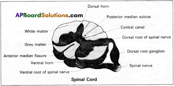
Draw a labelled diagram of the T.S. of the spinal cord of man.
Answer:
Question 2.
Distinguish between somatic and autonomic neural systems.
Answer:
| Somatic neural system | Myosin |
| 1. The senso’ry neurons conduct sensory impulses from the different somatic receptors to the CNS. | 1. The autonomic neurons are associ-ated with interoceptors. |
| 2. All these sensations are consciously perceived. | 2. These sensory signals are generally not continuously perceived. |
| 3. Somatic motor neurons innervate the skeletal muscles and produce voluntary movements. | 3. Autonomic motor neurons regulate the involutionary activities of the cardiac muscle, smooth muscle and glands. |
| 4. Acetyl choline is the neurotrans-mitter. | 4. Acetyl choline (or) norepinephrine is neurotransmitter. |
Question 3.
Give an account of the retina of human eye.
Answer:
Retina is the inner coat of the eye. It consist of a pigmented epithelium and a neural portion. The pigmented epithelium is a sheet of melanin containing epithelial cells. The neural portion has three layers namely photoreceptor Iaydr, bipolar cell layer and ganglion cell layer.
Photoreceptor layer consist of rods and cones. Rods contain a protein called rhodopsin. Rods are concerned with dim light. Cones contain a visual pigment called iodopsin and they are important in daylight vision and colour vision. There are three types of cones and are response to red, green and blue colours.
The centre of the posterior portion of the retina is called yellow spot. A depression present in the yellow spot is called ‘Forea’ contractile and it contains only cones. Forea is responsible for sharp vision. The region of retina which is devoid of rods and cones is known as blind spot (or) optic disc, which form the optic nerve called 2nd cranial nerve.
![]()
Question 4.
Give an account of synaptic transmission.
Answer:
A nerve impulse is transmitted from one neuron to another through junction called synapses.
There are two types of synapses. 1) Electrical synapses 2) Chemical synapses.
Electrical synapses :
These synapses are electrically conductive links between two neurons and are also called “gap junctions”. Impulses transmission across an electrical synapses is always faster than that across a chemical synapses.
Chemical synapses :
Chemicals called neuro transmitters are involved in the transmission of impulses at those synapses. When an impulse arrives at the axon terminal, it depolarizes the membrane opening voltage gated calcium channels. Calcium ions stimulate the release of neurotransmitters in the cleft by exocytosis. The released neurotransmitters bind to their specific receptors, present on the post synaptic membrane.
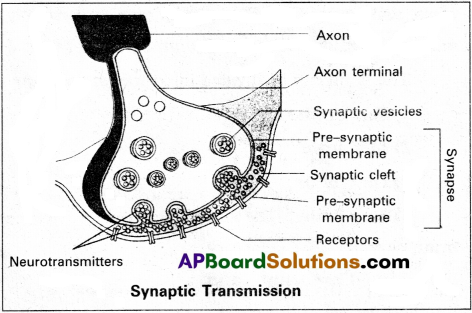
The post synaptic membrane has ligand gated channels. They are ion channels which respond to chemical signals, rather than to changes in the membrane potential. The entry of ions can generate a new potential in the post synaptic neuron. The new potential developed may be either excitatory (or) inhibitory.
Excitatory post synaptic potentials cause depolarisation, where as inhibitory post synaptic potentials cause hyper polarisation of post synaptic membrane.
Question 5.
List out the differences between Sympathetic and Parasympathetic neural system in man.
Answer:
| Sympathetic neural system | Parasympathetic neural system |
| 1. SNS originates in the thoracic and lumbar regions of the spinal cord. | 1. PNS originates in the cranial region of the brain and the sacral region of the spinal cord. |
| 2. Its ganglia are linked up to form a chain. | 2. Its ganglia remain isolated. |
| 3. Preganglionic fibres are short and the postganglionic fibres are long. | 3. Preganglionic fibres are long and the postganglionic fibres are short. |
| 4. Norepinephrine is produced at the terminal ends of the post-ganglionic fibres at the synapses on the effectors organ. Hence the system is called adrenergic’ usually. | 4. Acetycholine is produced at the ter-minal ends of the postganglionic fi-bres at the effector organ. Hence the system is called cholinergic’ usually. |
| 5. Active during stressful conditions preparing the body to face them. | 5. Active during relaxing times, restor-ing normal activity after stress. |
| 6. The-overall effect is excitatory and stimulating. | 6. The overall effect is inhibitory. |
Long Answer Questions
Question 1.
Give a brief account of the structure and functions of the brain of man.
Answer:
Brain is the site of information, processing and control. It is protected in the cranial cavity and covered by three cranial meninges namely duramater (outer layer), arachnoid mater (thin middle layer) and piamater (inner layer).
The brain can be divided into three major parts called
- Fore brain
- Mid brain
- Hind brain.
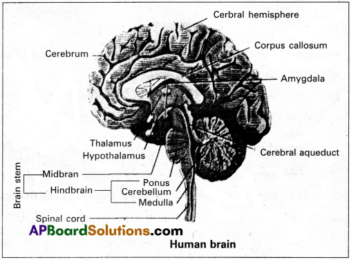
1) Fore brain :
The fore brain consists of i) Olfactory bulb ii) Cerebrum and iii) Dience-phalon.
i) Olfactory bulb :
Which receives impulses pertaining to smell from the Olfactory epithe-lium.
ii) Cerebrum :
Cerebrum forms the major part of the human brain. A deep cleft divides the cerebrum longitudinally into two halves, which are termed as the left and right cerebral hemispheres. The hemispheres are connected by a transverse, wide and flat bundle of myelinated fibres beneath the cortex, called corpus callosum. It brings the coordination between the left and right sides of the hemispheres. The surface of the cerebral cortex shows many folds and grooves. The folds are called gyri, the deepest and shallower grooves between folds are called fissures and sulci respectively.
The cerebral cortex contain three functional areas called
a) Sensory areas : receive and interpret the sensory impulses.
b) Motor areas : which control volutntary muscular movements.
c) Association areas : which are neither clearly sensory nor motor in function, they deal integrative functions, such as memory and communications.
The cerebral medulla consist of mostly myelinated axons. Each cerebral hemisphere of the cerebrum is divided into four lobes namely frontal, parietal, temporal and occipital lobes.
iii) Diencephalon :
It contains three main parts namely, a) Epithalamus, b) Thalamus and c) Hypothalamus.
a) Epithalamus :
It is the roof of the diencephalon. It is axon nervous part which is fused with the pia matter to form the anterior choriod plexus. The epithelium of the epithalamus forms a pineal stalk, which ends in a rounded structure called pineal body.
b) Thalamus :
It lies superior to the mid brain. It is the major coordination centre for sensory and motor signalling.
c) Hypothalamus :
It lies at the base of the thalamus. The hypothalamus forms a funnel-shaped downward extension called infundibulum, connecting the hypothalamus with the pituitary gland. It also contains a group of neuro-secretory cells, which secrete hormones called hypothalamic hormones.
Hypothalamus controls and integrates the activities of the autonomous nervous system and it has osmoregulatory, thermoregulatory, thirst, feeding the satiety centres.
Limbic system :
The inner part of cerebral hemisphere and group of associated structures forms limbic system. Limbic system along with hypothalamus is involved in the regulation of sexual behaviour and expression of emotional reactions.
2) Mid brain :
Mid brain is located between the thalamus of the fore brain and pons varolii of hind brain. The ventral portion of mid brain consists of a pair of longitudinal bands of nervous tissues called cerebral peduncles. The dorsal portion of the mid brain consists of four lobes called corpora quadrigmina. The two larger anterior lobes are called superior colliculi, which are concerned with visual function. The smaller posterior lobes are called inferior colliculi and are concerned with auditory functions.
3) Hind brain :
The hind brain comprises of cerebellum, pons varolii and medulla oblongata.
i) Cerebellum :
It is the second largest part of the brain. It consists of two cerebellar hemispheres and a central vermis. Each cerebellar hemisphere consists of three lobes namely anterior, posterior and floccular lobes. It has a branching tree like core of white matter called arbor vitae.
ii) Pons Varolii :
It consists of nerve fibres which form a bridge between the two cerebellar hemispheres. It is a relay station between the cerebellum, spinal cord and the rest of the brain. Pons has the pneumotaxic centre as it regulates the amount of air a person can take in each time.
iii) Medulla Oblongata :
It is the posterior part of brain. It extends from the Pons Varolii above and continuous with the spinal cord below. Medulla includes cardiovasicular, and respiratory centers, the centers for swallowing, vomiting, coughing, sneezing and hiccupping. The mid brain, pons and medulla Oblongata are collectirel referred to as brain stem.
![]()
Question 2.
Explain the transmission of nerve impulse through a nerve fibre with the help of suitable diagrams.
Answer:
Nerve impulse is the combination of mechanical, chemical (or) electrical disturbances occur in neuron because of stimulus. The propagation of a impulse along nerve fibre is called transmission. In this process both physical and chemical changes are involved. The entire process is divided into stimulation, excitation, conduction and response.
Resting membrane potential :
The resting membrane potential exists because of a small buildup of negative ions in the axoplasm along the inside of the membrane and an equal buildup of positive ions in the extra cellular fluid along the outer surface of the membrane. Such a Separation of positive and negative electrical chafges is a form of potential energy. In neurons, the resting membrane potential ranges from -40 to -90 mV. A typical value is-70 mV.
At resting phase, the axolemma is polarized. If the inner side becomes less negative, it is said to be depolarized. If the inner side becomes more negative, it is said to be hyperpolarized. During the resting phase the activation gates of sodium are closed, the inactivation gates of sodium are open and the activation gates of potassium are closed.
Sodium-potassium pump : Sodium and potassium ions diffuse inwards and outwards, respectively, down their concentration gradients through leakage channels. Such a movement of ions, if unchecked, would eventually disturb the resting membrane potential. These flows of ions are offset by sodium-potassium pumps (Na+/K+ ATPases) present in the axonal walls. These pumps expel three Na+ ions for each two K+ ions imported. As these pumps remove more positive charges from the axoplasm than they bring into it, they contribute to the negativity of the resting membrane potential i.e.,-70mv.
Depolarization (Rising phase):
When a nerve fibre is stimulated, the plasma membrane becomes more permeable to Na+ ions than to K+ ions as the activation and inactivation voltage gates of sodium open and activation voltage gates of potassium close. As a result the rate of flow of Na+ into the axoplasm exceeds the rate of flow of K+ to the ECF. Hence, the axolemma is positively charged inside and negatively charged outside. This reversal of electrical charge is called “depolarization”.
Outer face of the point which is adjacent to the site of depolarization remains positively charged. The electrical potential difference between these two areas is called “action potential”. An action potential occurs in the membrane of the axon of a neuron when depolarization reaches a certain level called ‘threshold potential1 (-55 mV). The particular stimulus which is able to bring the membrane potential to threshold is called ‘threshold stimulus’.
Repolarization (Falling phase) :
As the wave of depolarization passes away from its site of origin to the adjacent point, the activation gates of sodium remain open, inactivation gates of sodium close and activation gates of potassium open at the site of origin of depolarization. As a result the influx of Na+ ions into the axoplasm from the ECF is checked and ‘efflux’ of K+ ions occurs, which leads to the returning of axolemma to the resting state (exit of potassium ions causes a reversal of membrane potential to negative inside). This is called ‘repolarization’.
Hyperpolarization (Undershoot):
The repolarization typically goes more negative than the resting potential to about -90 mV This is called ‘hyperpolarization’. This occurs because of the increased K+ permeability that exists while voltage gated K+ channels are open activation and inactivation gates of Na+ channels remain closed. The membrane potential returns to its original resting state as the K+ channels close completely. As the voltage falls below the -70 mV level of the resting state, it is called ‘undershoot’.
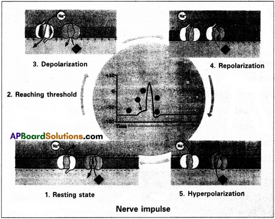
The refractory periods :
The period of time after an action potential begins during which the neuron cannot generate another action potential in response to a normal threshold stimulus is called the ‘refractory period’. There are two kinds of refractory periods, namely the absolute refractory period and the relative refractory period. During the absolute refractory period, even a very strong stimulus cannot initiate a second action potential. The relative refractory period is the time during which a second action potential can be initiated by a larger than normal stimulus.
Conduction speed:
The conduction speed of a nerve impulse depends on the diameter of the axon: the greater the axon’s diameter, the faster the conduction. In a myelinated axon, the voltage-gated Na+ and K+ channels are concentrated at the nodes of Ranvier. As a result, the impulse ‘jumps’ from one Ranvier’s node to the next, rather than travelling the entire length of the nerve fibre. This mechanism of conduction is called Saltatory conduction. Saltatory conduction is faster (in myelinated fibres) than continuous conduction (in nonmyelinated fibres).