Andhra Pradesh BIEAP AP Inter 1st Year Zoology Study Material Lesson 7 Type Study of Periplaneta Americana (Cockroach) Textbook Questions and Answers.
AP Inter 1st Year Zoology Study Material Lesson 7 Type Study of Periplaneta Americana (Cockroach)
Very Short Answer Type Questions
Question 1.
Why do you call cockroaches a pest?
Answer:
The cockroach is a common household pest that contaminates our food with its excreta and can transmit a number of diseases.
Question 2.
Name the terga of thoracic segments of cockroaches.
Answer:
Tergum of prothorox is Pronotum.
The tergum of mesothorax is Mesonotum.
The tergum of metathorax is Metanotum.
Question 3.
What are the structures with which cockroach walks on smooth surfaces and on rough surfaces respectively?
Answer:
The claws and the arotium help in locomotion on rough surfaces whereas planulae are useful on smooth surfaces.
Question 4.
Why is the head in cockroach called hypognathous?
Answer:
It lies hinging almost a right angles to the body with the posterior wider part upwards and the mouth parts directed downwards.
Question 5.
How is a tripod formed With reference to locomotion in cockroach?
Answer:
Tripod is formed by foreleg and hind leg of one side middle leg of other side. The fore leg and hind leg of the tripod kept on the ground, pull and push the body, while the middle leg acts as a pivot.
![]()
Question 6.
Name the muscles that help in elevating and depressing the wings of a cockroach.
Answer:
Wings are elevated by the contraction of dorsoventral muscles. Contraction of the dorsa longitudinal muscles depresses the muscles.
Question 7.
Name the different blood sinuses in cockroach.
Answer:
The three sinuses of haemocoel are known as
Pericardial haemocoel/Dorsal sinus
Perivisceral haemocoel/middle sinus
Perineural haemocoel/ventral sinus.
Question 8.
How are the fat bodies similar to the liver of the vertebrates?
Answer:
Fat bodies have many cells that are similar to the liver of the vertebrates in certain functions, namely
Trophocytes (store food)
Mycetocytes (contain symbiotic bacteria)
Oenocytes (secrete lipids)
Urate cells (store uric acid)
Question 9.
Which part of the gut secretes the peritrophic membrane in cockroach?
Answer:
Peritrophic membrane is secreted by the funnel like stomodel valve of the gizzard of midgut.
Question 10.
In which part of the gut of cockroach, water is reabsorbed?
Answer:
Rectum reabsorbes the water in cockroach.
Question 11.
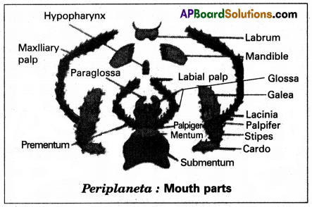
Write the names of mouthparts in cockroach that help in biting and tasting the food.
Answer:
Mandibles helps in biting and labrum helps in tasting the food.
Question 12.
What are alary muscles?
Answer:
A services of paired triangular muscles that are present in the dorsal and ventral diaphragm.
Question 13.
What is haemocoel?
Answer:
In cockroach blood (or) haemolymph flows freely with in the body cavity (or) haemocoel.
Question 14.
The three sinuses in a cockroach are not equal in size. Why?
Answer:
The middle sinus is very large as it contains most of the viscera. The dorsal and ventral sinuses are small as they have only heart and nerve cord.
Question 15.
Why is the blood of Periplaneta called haemolymph?
Answer:
The blood of periplaneta is colourless and it consists of fluid plasma and blood cells. Hence blood of periplaneta is called haemolymph.
![]()
Question 16.
What is the function of haemocytes found in the blood of Periplaneta?
Answer:
Haemocytes are phagocytic in nature. These are large in size and can ingest foreign particles.
Question 17.
Why does not the blood of Periplaneta help in respiration?
Answer:
Due to the absence of respiratory pigment the blood of cockroach can’t carry oxygen to different tissues.
Question 18.
Write important functions of blood in Periplaneta.
Answer:
1. It absorbs digest food from alimentary canal and distributes it to the rest of the body.
2. It transports secretions of the ductless glands to the target organs.
Question 19.
How many spiracles are present in cockroach? Mention their locations.
Answer:
Ten pairs of spiracles are present in cockroach.
Location: First two pairs of spiracles are present in the thoracic segments, remaining eight pairs present in first eight abdominal segments. Spiracles are located in the pleura of their respective segment.
Question 20.
What are trichomes? Write their functions.
Answer:
Trichomes are small hair-like structures of spiracles.
Function: Filtering the dust particles.
Question 21.
Why is the respiratory system of cockroaches called polytheistic and holocaustic systems?
Answer:
The spiracles of cockroaches are more in number (10 pairs) and all are functional so the respiratory system of cockroaches is called polytheistic and holocaustic systems.
Question 22.
What is intima?
Answer:
A cuticle layer that forms the inner layer of trachea is called intima.
Question 23.
During inspiration which spiracles are kept open and which are kept closed?
Answer:
Thoracic spiracles are kept open and the abdominal spiracles are kept closed.
Question 24.
Which factors regulate the opening of the spiracles?
Answer:
Opening and closing of spiracles is influenced by CO2 tension in haemolymph and oxygen tension in the trachea.
Question 25.
Inspiration in cockroach is a passive process and expiration is an active process. Justify?
Answer:
As air is drawn in due to the relaxation of the muscle inspiration is a “passive process”. Expiration involves the contraction of muscles, so it is described as active process.
![]()
Question 26.
The nitrogenous wastes in Periplaneta are removed from the body through alimentary canal. Why?
Answer:
Malphigian tubules collect nitrogenous acts from the body parts and releases into alimentary canal. So these nitrogenous wastes get mixed with facel matter and sent out through anus.
Question 27.
How does the cuticle of a cockroach help in excretion?
Answer:
Some nitrogenous waste materials are deposited on the cuticle and eliminated during moulting.
Question 28.
How do fat bodies help in excretion?
Answer:
Urate cells present in these bodies are associated with excretion in a way. Those cells absorb and store uric acid.
Question 29.
What is ‘storage excretion’?
Answer:
Urate cells present in the fat bodies absorb and store uric acid throughout life. This is called “storage excretion”.
Question 30.
Which structure of the cockroach acts as a sensory and endocrine centre?
Answer:
The brain of the cockroach acts as the sensory and endocrine centre.
Question 31.
Distinguish between scolopidia and sensillae.
Answer:
| Scolopidia | Sensillae |
| Sub-cuticular units of mechano receptors of chordotonal organs. | Units of cuticular receptors and chemoreceptors. |
Question 32.
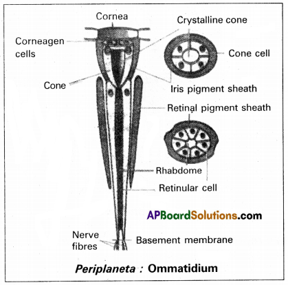
How is the ommatidium of cockroaches different from that of diurnal insects?
Answer:
Retinulae are present deep below the vitrallae and crystalline cone. The retinal sheath is absent.
Question 33.
Which of the abdominal ganglia is the largest and why?
Answer:
6th abdominal ganglia are the largest of all the abdominal ganglia because it is formed by the fusion of the ganglia of the 7th, 8th, 9th & 10th abdominal segments.
Question 34.
Name the structural and functional unit of the compound eye of the cockroach. How many such units are present in a single compound eye?
Answer:
Each compound eye is composed of about 2000 functional units called ommatidia.
Question 35.
Why is the brain called the principal sensory centre in cockroaches?
Answer:
The brain receives sensory impulses from various mouth parts & compound eye. Hence the brain is the principally a sensory centre.
![]()
Question 36.
Distinguish between apposition image and superposition image.
Answer:
| Apposition | Superposition |
| 1. These images are formed in diurnal insects. | 1. These images are formed in nocturnal insects. |
| 2. Mosaic image is formed. | 2. Overlapping (Blurred) image formed. |
| 3. Vision is mosaic. | 3. Vision is not clear. |
Question 37.
List out the characters that help in understanding the difference between male and Female cockroaches.
Answer:
| Male | Female |
| 1. Eight terga are not visible. | 1. Both eighth & ninth terga are not visible. |
| 2. Nine sterna are visible. | 2. Only seven sterna are visible. |
| 3. Anal styles are present. | 3. Anal styles are absent. |
Question 38.
What is the function of the mushroom gland in cockroaches?
Answer:
A characteristic mushroom-shaped gland is present in the 6th and 7th abdominal segments which functions as an accessory reproductive gland.
Question 39.
Compare the utriculi majors and utriculi breviores of the mushroom gland functionally.
Answer:
- Utriculi majores forms the inner layer of the spermatophore.
- Utriculi breviores nourish the sperms.
Question 40.
What are Phallomeres?
Answer:
Surrounding the male genital opening there are chitinous and asymmetrical structures called phallic organs/phellomeres/gonapophyses which help in copulation.
Question 41.
What is gona Pophyses?
Answer:
Surrounding the male genital opening there are chitinous and asymmetrical structures called phallic organs/phellomeres/gonapophyses which help in copulation.
Question 42.
How is the colleterial gland helpful in the reproduction of Periplaneta?
Answer:
A pair of branched colleterial glands is present behind the ovaries. These glands open into the genital pouch separately. Secretions of the two collateral glands form a hard egg case called Ootheca.
Question 43.
What is paurometabolous development?
Answer:
Gradual development (metamorphosis) through nymph stages is called “parametabolous development”.
Ex: Periplaneta
Short Answer Type Questions
Question 1.
Draw a neat labelled diagram of the mouthparts of cockroaches.
Answer:
Question 2.
Describe the physiology of digestion in cockroaches.
Answer:
Food collection: The cockroach is an omnivorous insect. It feeds on all types of organic matter.
Digestion: After swallowing, the food passes through the pharynx and oesophagus and reaches the crop. In the crop, food is mixed with digestive juices that are regurgitated into it through the grooves of the gizzard. Hence, most of the food is digested in the crop. The partly digested food is filtered by the bristles of the gizzard and later it passes through the stomodeal valve into the ventricular.
The enzyme amylase of the salivary juice converts starches into disaccharides. Invertase or sucrase digests sucrose into glucose and fructose. Maltose converts maltose into glucose. The enzyme lipase digests lipids into fatty acids and glycerol. Proteases digest proteins into amino acids. The cellulose of the food is digested by the enzyme cellulose secreted by the microorganisms present in the hindgut of cockroaches. Cellulose is converted into glucose.
In the ventriculus, the digested food is absorbed. The undigested food is passed into the ileum, and colon and then reaches the rectum, where water is reabsorbed by rectal papillae. Then the remaining material is finally defecated as dry pellets, through the anus.
![]()
Question 3.
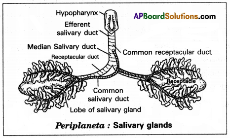
Draw a neat labelled diagram of the salivary apparatus of cockroaches.
Answer:
Question 4.
Describe the structure and function of the heart in Periplaneta.
Answer:
Heart: The heart lies in the pericardial hemocoel or dorsal sinus. It is a long muscular, contractile tube found along the mid-dorsal line, beneath the terga of the thorax and abdomen. It consists of 13 chambers. Every chamber opens into the other present in front of it. Three of the thirteen chambers are situated in the thorax and ten in the abdomen. Its posterior end is closed while the anterior end is continued forward as the anterior aorta. On the posterior side of each chamber, except the last, there is a pair of small apertures called ‘Ostia’ one on each side. Ostia have valves that allow the blood to pass only into the heart from the dorsal sinus.
Question 5.
Describe the process of blood circulation in Periplaneta.
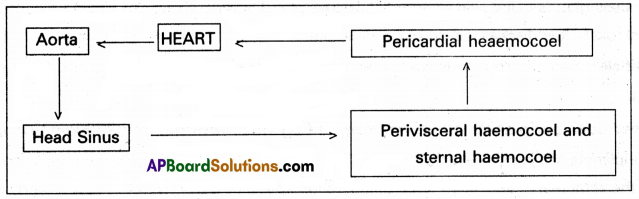
Answer:
The blood flows forward in the heart by the contractions of its chambers. At the anterior end of the heart, the blood flows into the aorta and from there it enters the sinus of the head. From the head sinus, the blood flows into the perivisceral and sternal sinuses. On contraction of the alary muscles, the pericardial septum is pulled down. This increases the volume of the pericardial sinus. Hence blood flows from the perivisceral sinus into the pericardial sinus through the apertures of the pericardial septum. On relaxation of the alary muscles, the pericardial septum moves upwards to its original position. This forces the blood, to enter the chambers of the heart through the Ostia from the pericardial sinus.
Question 6.
How do contraction and relaxation of alary muscles help in circulation?
Answer:
The blood flows forward in the heart by the contractions of its chambers. At the anterior end of the heart, the blood flows into the aorta and from there it enters the sinus of the head. From the head sinus, the blood flows into the perivisceral and sternal sinuses. On contraction of the alary muscles, the pericardial septum is pulled down. This increases the volume of the pericardial sinus. Hence blood flows from the pericardial septum. On relaxation of the alary muscles, the pericardial septum moves upwards to its original position. This forces the blood, to enter the chamber of the heart through the Ostia from the pericardial sinus.
Question 7.
What are the different excretory organs in Periplaneta? Describe the process of excretion in detail.
Answer:
The structures associated with excretory function are Malpighian tubules, Fat bodies, uricase glands, Nephrocytes, and Cuticles.
Malpighian tubule: The malpighian tubules are long, unbranched yellowish tubules, attached at the extreme anterior end of the hindgut, lying freely in the hemolymph, but do not open into it being bliand at the free ends. They are 100-150 in number arranged in 6-8 bundles, each bundle having 15-25 tubules. Each tubule is lined by a single layer of glandular epithelium with a brush border on the inner surface. The ‘distal portion’ of the tubule is secretory and the ‘proximal part’ is absorptive in nature.
The glandular cells of the malpighian tubules absorb water salts, CO2, and nitrogenous wastes from the hemolymph and secrete them into the lumen of the tubules. The cell of the proximal part of the tubules reabsorbs water and certain inorganic salts. By the contraction of the tubules, urine is pushed into the ileum. More water is reabsorbed from it when it moves into the rectum and almost solid uric acid is excreted along with faecal matter.
The removal of nitrogenous waste material through the alimentary canal helps in the complete reabsorption of water from the wastes and the formation of dry uric acid. It is an adaptation for the conservation of water as it is very important in terrestrial organisms.
Fat bodies: Fat body is a lobed white structure. Urate cells present in these bodies are associated with excretion in a way. These cells absorb and store uric acid throughout life. This is called storage excretion as they remain stored in the cells of the corpora adipose.
Uricose glands: Uric acid is stored in uriosa gland or utriculi majority of the mushroom gland in male cockroaches. It is discharged during copulation.
Cuticle: Some nitrogenous waste materials are deposited on the cuticle and eliminated during moulting.
![]()
Question 8.
How does Periplaneta conserve water? Explain it with the help of excretion in it.
Answer:
Periplaneta can conserve water by following methods. The removal of Nitrogenous waste material through the alimentary canal helps in the complete reabsorption of water from the wastes and the formation of dry uric acid. It is an adaption for the conservation of water as it is very important in terrestrial organisms.
Cuticle: Some nitrogenous waste materials are deposited on the cuticle and eliminated during moulting.
Question 9.
Draw a neat and labelled diagram of Ommatidium.
Answer:
Question 10.
How can you identify the male and female cockroaches? Explain it describing the chief structures of the external and internal genitalia.
Answer:
Periplaneta is dioecious or unisexual and both the sexes have well-developed reproductive organs. Sexual dimorphism is evident both externally and internally. The female is different from the male in respect of short and broad abdomen, presence of blood pouches, and absence of anal styles.
The eighth tergum in the male and both the eighth and ninth terga in the female are not visible. In the male ninth sterna are visible, whereas in the female only the seventh sterna are visible. The seventh, eighth, and ninth sterna together form a broad pouch.
The posterior end of the abdomen is a pair of anal cerci, a pair of anal styles and gonophophyses in the males, and cerci are jointed and arise from the lateral side of the tenth tergum and are found in both sexes. The anal styles are without joints and arise from the ninth sternum. But seen only in the males. The gonopophysis are small chitinous processes arising from the ninth sternum in males and the eighth, and ninth sterna in females. They are the external genital organs.
Question 11.
Describe the male reproductive system of cockroaches.
Answer:
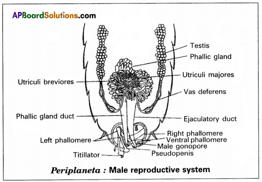
The male reproductive system consists of a pair of testes. These are elongated and lobed structures lying on each lateral side in the fourth to sixth abdominal segments. They are embedded in the fat bodies. From the posterior end of each testis, there starts a thin duct, the vas deferens.
The two vasa deferentia run backward and inwards to open into a wide median duct, the ductus ejaculatory in the seventh segment. A characteristic mushroom-shaped gland is present in the 6th and 7th abdominal segments which functions as an accessory reproductive gland.
The gland consists of two types of tubules:
- Long slender tubules, the utriculi majors, or peripheral tubules.
- Short tubules, the utriculi breviores, and secretion of utriculi majors form the inner layer of the spermatophore while the utricular breviores nourish the sperms. These tubules open into the anterior part of the ejaculatory duct.
Question 12.
Describe the female reproductive system of cockroaches.
Answer:
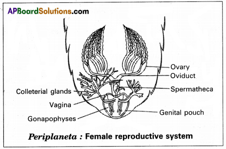
The female reproductive system of Periplaneta consists of a pair of ovaries a pair of oviducts vagina, spermathecal papilla, colleterial glands, and ovaries.
A pair of large ovaries lie laterally in 2 to 6 abdominal segments. They are light yellow in colour surrounded by fat bodies. Each ovary consists of eight tubules called ovarian tubules or ovarioles. Each ovariole consists of a tapering anterior filament called germarium and a posterior wider vitellarium. The germarium contains various stages of developing ova and the vitellarium contains mature ova with the yolk. The tapering ends of the ovarioles of each ovary unite to form a single thread that attaches to the dorsal body wall.
The ovarioles, at their posterior end, unite to form a short wide oviduct. The oviducts unite to form a very short median vagina. The vertical opening of the vegina is called the female genital pore. It opens into a large genital pouch on the eighth sternum. A spermatheca or receptaculum seminis, consisting of a left-sac like and a right filamentous caecum, is present in the 6th segment which opens by a median aperture on a small spermathecal papilla in the dorsal wall of the genital pouch on the ninth sternum. In a fertile female, the spermatheca contains spermatophores, obtained during copulation.
A pair of branched colleterial glands is present behind the ovaries. These glands open into the genital pouch separately, just above the spermathecal aperture. Secretion of the two collateral glands forms a hard egg case called ootheca around the eggs.
Long Answer Type Questions
Question 1.
Describe the digestive system of cockroaches with the help of a neat labelled diagram.
Answer:
The digestive system of cockroaches consists of an alimentary canal and the associated glands. The preoral cavity surrounded by the mouth parts is present in front of the mouth. The hypopharynx divides into two chambers called cibagium (anterior) and salivarium (posterior).
Alimentary canal: The alimentary canal of cockroaches is a long tube and is coiled in some places. It extends between the mouth and the anus. It is divided into three regions namely the foregut of stomodaeum, midgut or mesenteron, and hindgut is internally lined by ectoderm. The mesenteron is lined by the endodermal cells.
Forgut or stomodaeum: The foregut includes the pharynx oesophagus, crop, and gizzard. It is internally lined by a chitinous cuticle. The mouth opens into the pharynx, which in turn leads into a narrow tubular oesophagus. The oesophagus opens behind into a thin-walled distensible sac called a crop. The crop serves as a reservoir for storing food. Its outer surface is covered by a network of tracheae.
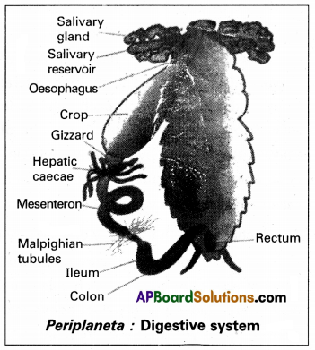
Behind the crop, there is a thick-walled muscular proven- triculus or gizzard. The chitinous inner living of the gizzard has six powerful teeth, which form an efficient grinding apparatus. Behind each tooth is a hairy pad, which bears backwardly directed bristles. Among these plates, food is thoroughly ground into fine particles. These food particles are filtered by the bristles. The gizzard thus acts both as a grinding mill and also as a sieve. There is a membranous projection of the gizzard into the mesenteron in the form of a funnel called a stomodeal valve. This valve prevents the entry (regurgitation) of food from the mesenteron back into the gizzard.
Midgut (mesenteron or ventriculus): The midgut is a short and narrow tube behind the gizzard. It is also called mesenteron or ventriculus. Between the ventriculus and the gizzard, arising from the ventriculus there are six to the eighth finger-like diverticula called nepatic caecae. They are helpful in the digestion and absorption of digested food materials. Ventriculus is functionally divided into an anterior secretory part and a posterior absorptive part.
The secretory part of the ventriculus has many gland cells and it secretes several enzymes. The ‘bolus’ of food in the mesenteron is enveloped by a chitinous and porous membrane called a peritrophic membrane, which is secreted by the funnel-like stomodeal valve of the gizzard. Digested food is absorbed into the food through the peritrophic membrane in the posterior absorptive region of the ventriculus. The peritrophic membrane protects the wall of the ventriculus from hard food particles in the food. The opening of the ventriculus into the hindgut is controlled by a sphincture muscle. It prevents entry of undigested food from the hindgut into the midgut.
Hindgut or proctodaeum: The hindgut is a long coiled tube, consisting of three regions namely the ileum, colon, and rectum. It is internally lined by the chitinous cuticle. The ileum that lies behind the mesenteron is a short tube. Six bundles of fine yellow, blind tubules called Malpighian tubules open into the ileum near the junction of mesenteron and ileum. Malpighian tubules are excretory in function. The ileum collects uric acid from the malpighian tubules and undigested food from the mesenteron. The ileum opens behind into a long coiled tube called the colon. The colon leads into a short and wide rectum which opens out through the anus. The rectum bears on its inner side six longitudinal chitinous folds called rectal papillae. They are concerned with the reabsorption of water from undigested food.
Digestive gland: The digestive glands associated with the alimentary canal of cockroaches are salivary glands, hepatic caecae, and glandular cells of the mesenteron.
Salivary glands: There is a pair of salivary glands attached to the ventrolateral sides of the crop, one on each side. Each salivary gland has two lobes. Each lobe of the salivary gland has many lobules called acini. Each acinus is a group of secretory cells called zymogen cells with a small ductule. The ductules of both the lobes of a salivary gland unite to form a common salivary duct on each side.
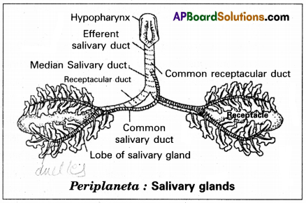
The two common salivary ducts are joined to form the median salivary duct. Between the two lobes of a salivary gland on each side is a sac called the salivary receptacular duct or common reservoir duct. The midious salivary duct opens into the common receptacular duct. Later these two form an efferent salivary duct. The efferent salivary duct opens at the base of the hypopharynx. Acinar cells secrete saliva, which contains a starch digesting enzymes such as amylase.
Hepatic caecae: The hepatic caecae are also termed midguts caecae. They contain secretory and absorptive cells.
Glandular cells of the mesenteron: The glandular cells of the mesenteron secrete enzymes such as maltase, invertase, proteases, and lipase.
![]()
Question 2.
Describe the blood circulatory system of Periplaneta in detail and draw a neat and labelled diagram of it.
Answer:
The circulatory system helps in the transportation of digested food, hormones, etc., from one part to another in the body. Periplaneta has an open type of circulatory system as the blood or hemolymph, flows freely within the body cavity or hemocoel, Blood vessels are poorly developed and open into spaces) Visceral organs located in the hemocoel are bathed in the blood. The three main parts associated with the blood circulatory system of Periplaneta are the hemocoel, heart, and blood.
Haemocoel: The haemocoel of cockroaches is divided into three sinuses by two muscular, horizontal membranes called dorsal diaphragm or pericardial septum and ventral diaphragm. Both diaphragms have pores. There is a series of paired triangular muscles called alary muscles. Every segment has one pair of these muscles situated on the lateral sides of the body. These are attached to the pericardial septum by their broad bases and to the terga by their broad bases and to the terga by their pointed ends or apices. The three sinuses of the haemocoel are known as pericardial haemocoel or the dorsal sinus, the perivisceral haemocoel or the middle sinus, and sternal haemocoel or ventral sinus or perineural sinus. The middle sinus is very large as it contains most of the viscera. The dorsal and ventral sinuses are small as they have only the heart and nerve cords, respectively.
Heart: The heart lies in the pericardial haemocoel or dorsal sinus. It is a long muscular, contractile tube found in a long mid-dorsal line, beneath the terga of the thorax and abdomen.
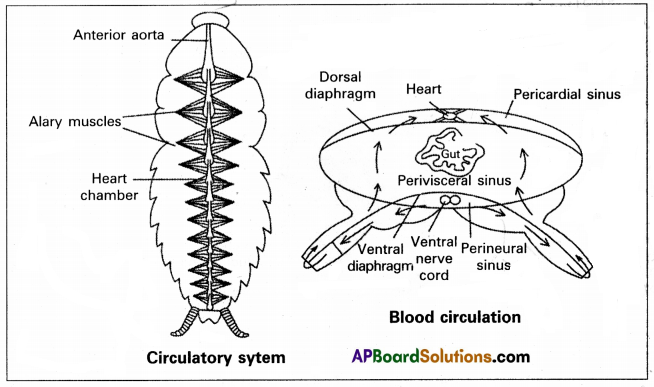
It consists of 13 chambers. Every chamber opens into the other present in front of it. Three of the thirteen chambers are situated in the thorax and ten in the abdomen. Its posterior end is closed while the anterior end is continued forward as the anterior aorta. At the posterior side of each chamber, except the last, there is a pair of small apertures called ‘Ostia’ one on each side. Ostia have valves that allow the blood to pass only into the heart from the dorsal sinus.
Blood: The blood of Periplaneta is colourless and is called haemolymph. It consists of a fluid called plasma and free blood corpuscles or haemocytes, which are phagocytic. The phagocytic, the phagocytes are large in size and can ‘ingest’ foreign particles such as bacteria. There is no respiratory pigment in the blood and so it plays no major role in respiration.
Question 3.
Describe the respiratory system of cockroaches with the help of neat and labelled diagrams.
Answer:
Due to the absence of respiratory pigment, the blood of cockroaches is colourless and it cannot carry oxygen to different tissues. Therefore a tracheal system is developed to carry the air directly to the tissues. The respiratory system of cockroaches consists of stigmata, tracheae, and tracheoles.
Stigmata or spiracles: The tracheal system communicates with the exterior by ten pairs of openings called stigmata or spiracles. The first two pairs of spiracles are present in the thoracic segments, one pair in the mesothorax and one pair in the metathorax. The remaining eight pairs of abdominal segments.
Spiracles are located in the pleura of their respective segments. The respiratory system in insects is classified on the basis of the number and nature of spiracles. The spiracles of cockroaches are polypneustic (as they are more than 3 pairs) and holopneustic (as all of them are functional). All spiracles are valvular and each of them is surrounded by a chitinous ring called peritreme. All spiracles bear small hair-like structures called trichomes to filter the dust particles.
Each spiracle opens into a small chamber called the atrium.
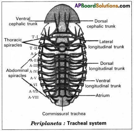
Tracheae: From the atrium of each thoracic spiracle several horizontal tracheae run inside. They join with each other in the thorax to form many tracheal trunks like dorsal cephalic, and ventral cephalic trunks and their branches. These branches enter all organs of the head. The thoracic region also contains lateral longitudinal trunks. The abdominal spiracles lead into the atria. From the atrium of each abdominal spiracle, three tracheal tubes arise. All these tracheal tubes on one side open into three separate longitudinal tracheal trunks. They are lateral dorsal and ventral longitudinal trunks. Lateral longitudinal trunks are the longest tracheal trunks. The three pairs of longitudinal tracheal trunks on both sides are interconnected by many commissural tracheae. From all the tracheal trunks several branches are given out, which enter different organs. All tracheal branches entering an organ end in a special cell called tracheoles cell.
The wall of the tracheae is made of three layers. They are an outer basement membrane, a middle one cell thick epithelium, and an inner layer of cuticle called the intima. The intima is produced into spiral thickening called taenidia. The taenidia keep the tracheae always open and prevent it from collapsing.
Tracheoles: The terminal cell of the trachea is called tracheoblast or tracheole cell. It has several intracellular tubular extensions called tracheoles. Tracheoles are devoid of intima and taenidia. They are formed of a protein called tracheal. Tracheolar fluid is present inside the tracheoles. The level of the tracheal fluid varies with the metabolic activity of the insect. It is more when the insect is inactive and completely reabsorbed into the tissues when the insect is more active. Tracheoles penetrate the cell and are intimately associated with mitochondria (to supply oxygen to them).
![]()
Question 4.
Describe the reproductive system of Periplaneta and draw neat and labelled diagrams of it.
Answer:
Periplaneta is dioecious, or unisexual and both the sexes have well-developed reproductive organs. Sexual dimorphism is evident both externally and internally. The female is different. The female is different from the male in respect of short and broad abdomen, presence of brood pouches, and absence of anal styles.
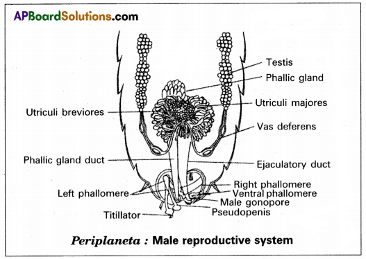
Male Reproductive system: The male reproductive system consists of a pair of testes. These are elongated and lobed structures lying on each lateral side in the fourth to sixth abdominal segments. They are embedded in the fat bodies. From the posterior end of each testis, there starts a thin duct, the vas deferens, the two vasa deferentia run backward and inwards to open into a wide median duct, the ductus ejaculators in the seventh segment. A characteristic mushroom-shaped gland is present in the 6th and 7th abdominal segments which functions as an accessory reproductive gland.
The gland consists of two types of tubules or i, long slender tubules, the utriculi majores or periphera tubules in short tubules, the utriculi breviores secretion of utriculi majores forms the inner layer of the spermatophore while that of utriculi breviores nourishes the sperms. These tubules open into the anterior part of the ejaculatory duct. The seminal vesicles are present on the ventral surface of the ejaculatory duct. These sacs store the sperms in the form of bundles called spermatophores. The ejaculatory duct is a muscular tube that extends posteriorly and opens at the gonopore or the male genital pore.
Female reproductive system: The female reproductive system of Periplaneta consists of a pair of ovaries, a pair of oviducts, vagina, spermathecae, spermathecal papilla, and colleterial glands.
Ovaries: A pair of large ovaries lie laterally in 2 to 6 abdominal segments. They are light yellow in colour surrounded by fat bodies. Each ovary consists of eight tubules called ovarian tubules or ovarioles. Each ovariole consists of a tapering anterior filament germarium and a posterior wider vitellarium. The germarium contains various stages of developing ova and the vitellarium contains mature ova with the yolk.
The tapering ends of the ovarioles of each ovary unite-to form a single thread that attaches to the dorsal body wall. The ovarioles, at their posterior end, unite to form a short wide oviduct. The oviduct unite to form a very short median vagina. The vertical opening of the vagina is called the female genital pore. It opens into a large genital pouch on the eighth sternum. A spermatheca or receptaculum seminis. Consisting of a left-sac-like and a right filamentous caecum is present in the 6th segment which opens by a median aperture on a small spermathecal papilla in the dorsal wall of the genital pouch on the ninth sternum.
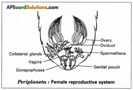
In a fertile female, the spermatheca contains spermatophores obtained during copulation. A pair of branched colleterial glands is present behind the genital pouch separately just above the spermathecal aperture, secretion of the two collateral glands forms a hard egg case called ootheca a round the eggs.
Three pairs of a plate-like chitinous structure called gonapophyses are present around the female genital aperture. These gonapophyses guide the ova into ootheca as ovipositors. These are the female external genitalia.